Difference of these two 12 lead monitoring?

DragonClaw

Emergency Medical Texan
2,116
363
83
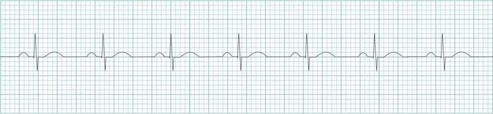
I don't really know how they're saying it's different. Or what to call them to distinguish the two. Is tracing the difference?

Also, I thought cardiac monitoring was a ALS procedure and EMTB wouldn't be doing this? I guess it depends on the medical director though?


4437

4438
 

Attachments

  • 1553817487120.png
    1553817487120.png
    247.6 KB · Views: 327

NPO

Forum Deputy Chief
1,831
897
113
One picture is showing limb leads. These go on the arms and legs (or as pictured, shoulders)

The other is showing chest leads. This is the part that's added that makes it a 12-lead. We hadd 6 stickers across the chest to look at those vectors.

It depends on local protocol, but typically if trained properly an EMT can place electrodes for 12 lead EKG acquisition. however, it is not typical for an EMT to be trained expected or authorized to interpret the EKG print out.
 

Aprz

The New Beach Medic
3,031
664
113
Yes, the tracings are different.

We usually start off with the bottom picture where it says step 4. We can put three to four wires "leads" on the patient, and we usually use these three to four leads to monitor the patient continuously. It might be documented as "cardiac monitoring", "3 lead ECG", or "4 lead ECG" depending on the number of wires used. We use the word "lead" interchangeably with the name of the actual wire (each wire is called a lead) and also each view of the heart the wire allows us to see, so even though there might be only 4 physical leads on the patient, it might produce 6 views/leads that we sometimes call "a quick 6".

If 3 physical leads are used only, usually with the monitor called a Philips MRx, then you'd put a red (left leg (LL)), white (right arm (RA)), and black lead (left arm (LA)) on the patient to monitor them. This would produce electrical views of the heart that we would call lead I, II, and III (those are roman numeral 1, 2, and 3, so we'd literally say lead "one", lead "two", and lead "three"). In the majority of places and training, lead II is used. Lead II is observed via the right arm white lead being considered a negative point, the red foot or red left leg lead being considered the positive point, and the black left arm lead being considered the ground lead that doesn't see or do anything or than be the ground. These negative and positive points would be in comparison to the overall average/mean electrical activity of the heart.

$_3.JPG
Philips MRx​

LEM1673A.jpg
Philips 3 leads​

ECG-Normal-sinus-rhythm-strip.jpg
ECG Strip, usually lead II unless said otherwise​

Other monitors like the Lifepak usually come with four physical wires, the same as Philips, but with a green right leg (RL) ground lead. To view lead II, you would only need to put the right arm white lead on (the negative point), the left leg/foot red lead (the positive point), and the green ground lead, which, would only show lead II, and no other lead could be seen. If you put all four wires/leads on the patient, it would produce a quick 6. With all four physical leads/wire on, you could see six leads/view lead I, II, III, aVR, aVL, and aVF.

tnw800-Lifepak%2015.jpg
Lifepak 15​

11111-000020_Detail.png
Lifepak 4 leads​

Image_4.png
Limb leads location on patients. Philips usually has green left leg lead attached to it's "12-lead" cables, but goes in the same location and serves the same purpose by being the ground lead​

These four wires, that we call the limb leads, show us the frontal/vertical view of the heart.

Limb%20Leads%20small%20version%20jpeg.jpg
Frontal view of the heart​

Anterior_Half.jpg
Quick six: I, II, III, aVR, aVL, and aVF​

This only shows us half the picture. So what we do to get a full view of the picture is put the precordial chest leads on the patient (step 5 in the initial post).

  • V1 is placed on the right side of the chest, 4th intercostal space, parasternal.
  • V2 is placed on the left side of the chest, 4th intercostal space, parasternal.
  • V3 is placed literally between lead V2 and V4. Usually people skip to lead V4 before placing lead V3 on.
  • V4 is placed on the left side of the chest, 5th intercostal, midclavicular.
  • V5 is placed on the left side of the chest, 5th intercostal, anterior axillary.
  • V6 is placed on the left side of the chest, 6th intercostal, midaxillary.

precordial-leads.jpg
Precordial chest leads​

M3527A-PM-ALS-Cable-3x3.jpg
Precordial chest leads with the green right leg ground lead used on Philips MRx​

These precordial chest leads give you a tranverse/horizontal view (as opposed to the frontal/vertical view of the limb leads).

precord_view.gif
Tranversed/horizontal view of the heart​

whylarge1.jpg
12 lead: Lead I, II, III, aVR, aVL, aVF, V1, V2, V3, V4, V5, and V6​

To monitor in these leads would be a little bit more work because I believe you need to have the limb leads on for them to work as well; These leads I believe use the very center of the limb leads as a second point to create a vector. The second point is called central terminus I believe. As you can see, it is kind of a pain to point on all these extra wires just to "monitor" the patient. That's why they aren't frequently used to monitor the patient, but just to get a full view of the heart. Sometimes people do like to use lead V1 to monitor for atrial arrhythmias because lead V1 rest right over the right atrium, and this could help you find something like "atrial flutter" which is a type of cardiac arrhythmia.

As paramedics, we generally do 3 leads or 4 leads to monitor the patient. When we suspect that there is an arrhythmia, ischemia "heart attack", electrolyte problem, we might do a 12-lead (limb leads and chest leads combine) to get the full view. Each lead serves different purposes that can help us identify cardiac arrhythmia (eg lead II, III, aVF, and V1 are useful in atrial tachycardia and flutter), localizing where there is heart damage in a heart attack (II, III, aVF are the inferior/bottom part of the heart. I, aVL, V5, and V6 are the left lateral part of the heart. V3 and V4 are the anterior/front part of the heart, or we can tell when there is damage to the back of the heart using the anterior part as a mirror.), or if there are electrolyte problems (particularly hyperkalemia/high potassium with tall T waves, usually I see it best in the precordial chest leads).

I would've kept this short and simple, but I feel like paramedic school usually lacks in this as well, or at least mine did. You will learn some or most of this in paramedic school, but I think it can be useful to know as an EMT working on an advance life (ALS) support unit with a paramedic why certain things may be done or why we might say something like "3 lead" vs "4 lead" or why the paramedic might not put all the wires on (my EMT will get critiqued by paramedics when she only puts the white, red, and green lead on, and they'll be like "You need to put on all of it on to work", which you actually don't). It might understand how a quick 6 might be useful, but also its disadvantage (you will miss anterior and posterior wall MIs "heart attacks" if you do not do a full 12-lead, but useful if you're afraid to give nitroglycerin to an inferior wall MI since a quick 6 does show the inferior leads "quickly").

Sorry for the novels. None of these pictures belong to me; I just stole them from Google.
 

Akulahawk

EMT-P/ED RN
Community Leader
4,938
1,341
113
There are other leads that are far less frequently used these days because 12 lead monitors are now very commonly used in the field. When I first got started, this wasn't the case. My Paramedic program did train us in 12 lead interpretation. They also knew that we were likely to NOT have 12-lead machines available to us quite yet, so we were also trained in how to set up monitors in different ways. The most common thing for us was to use Modified Chest Leads 1-6, of course we'd have to do them one at a time, as these approximated the V leads. In larger patients, I often chose (back then) to use MCL1 as I'd usually get less motion artifact. Aside from the physicians, I doubt many people in my ED know about the Lewis Lead...

In any event, the posts above have some good info. The 12-lead machines will typically have 4 leads you'd use for most cardiac monitoring. This does give you leads 1-3 and three additional leads (AVR, AVL, AVF) if you're inclined to use 'em. The monitors I use at work are 5 lead - same 4 limb leads and one "V" lead. Some field machines may have this option. If yours does, just make sure you place the V lead appropriately.

I, in my role as a Paramedic or RN, really don't care who places the leads on a patient as long as they're placed in their proper locations. If you're "my" EMT, I'm happy to train you where to place those leads, but I don't expect you to do any interpretation. If those leads aren't properly placed, the interpretation can't actually be trusted. The computer (or provider) doesn't know if the leads are poorly placed, so... bad data in = bad interpretation.
 

StCEMT

Forum Deputy Chief
3,052
1,709
113
You've already gotten a in depth answer, but to give you a short answer to your specific questions....they are different views of the heart. So since you are looking at the heart from different angles, yes, the tracings will be different.

Once you have an understanding of the electrical pathway of the heart, the tracings will make more sense. Combine understanding of the pathway with how the electrodes "see" the movement of electricity and how that is graphed out, you will understand why the patterns are the way they are. For example avR being inverted, and seeing a gradual increase in the R wave from V1 to V6.

This is where the A&P and other basic foundational knowledge that gets repeated so often comes into play.
 
OP
OP
DragonClaw

DragonClaw

Emergency Medical Texan
2,116
363
83
If I read correctly, there's not 12 wires though, so how does that generate 12 leads?

Also, does 1 special wire have to be the ground, or can any work (regardless of what wire generally goes where)?

If you need to use an AED (or manual), can you shock with the leads or will that fry the machine?
 

DrParasite

The fire extinguisher is not just for show
6,197
2,053
113
No, you don't shock with the 12 leads; you use different pads to deliver the electricity.

EMTs can apply the leads; however EMTs are not trained or educated enough to interpret the results.

If you just put the limb leads on, or apply the defib pads, all you will pick up is lead 2, which can show you some rhythm abnormalities, but if you want to see other views of the heart, you need to connect all the wires (and yes, 6+4 does not equal 12, but it's 12 different views of the heart)
 
OP
OP
DragonClaw

DragonClaw

Emergency Medical Texan
2,116
363
83
No, you don't shock with the 12 leads; you use different pads to deliver the electricity.

EMTs can apply the leads; however EMTs are not trained or educated enough to interpret the results.

If you just put the limb leads on, or apply the defib pads, all you will pick up is lead 2, which can show you some rhythm abnormalities, but if you want to see other views of the heart, you need to connect all the wires (and yes, 6+4 does not equal 12, but it's 12 different views of the heart)
I don't mean with. I mean, if you put the defibrillator pads on and shock, is it going to travel up the leads and make the monitor take a dirt nap? Or are they designed to withstand that electricity?
 
OP
OP
DragonClaw

DragonClaw

Emergency Medical Texan
2,116
363
83
And you're saying it's like permutations, with the possibilities of what's being interpreted by the monitor by comparing different leads?
 

mgr22

Forum Deputy Chief
1,660
820
113
And you're saying it's like permutations, with the possibilities of what's being interpreted by the monitor by comparing different leads?

Think of the different leads as cameras taking "pictures" of the heart from different angles.
 

DrParasite

The fire extinguisher is not just for show
6,197
2,053
113
Have you tried viewing more than Lead II after attaching all of the limb leads?
Not sure exactly what you are asking... you will only pick up one lead at at time, but the default is lead II; you can change the settings to pick up another lead. But every time I have seen someone want to see more than one lead, we just run a 12 lead. so I guess the answer is rarely?
 

mgr22

Forum Deputy Chief
1,660
820
113
Not sure exactly what you are asking... you will only pick up one lead at at time, but the default is lead II; you can change the settings to pick up another lead. But every time I have seen someone want to see more than one lead, we just run a 12 lead. so I guess the answer is rarely?

You had said "If you just put the limb leads on, or apply the defib pads, all you will pick up is lead 2." That sounded like you were unaware you could view more than lead 2 with limb leads. I was going to explain that to you and clarify it for the OP before you corrected yourself.
 

VFlutter

Flight Nurse
3,728
1,264
113
There are 10 electrodes, 3/4 of the Limb leads are Bipolar and are used to create 12 different views that we call "leads"
 

StCEMT

Forum Deputy Chief
3,052
1,709
113
Go back over your book and read about how all this works, you will answer a lot of your own questions. If something doesn't make sense, tell us what you've read/your understanding is and we can clarify from there.
 

E tank

Caution: Paralyzing Agent
1,580
1,430
113
Dang...I feel a little weird taking my shirt off at the pool....but...dude...
 

DesertMedic66

Forum Troll
11,273
3,452
113
I don't mean with. I mean, if you put the defibrillator pads on and shock, is it going to travel up the leads and make the monitor take a dirt nap? Or are they designed to withstand that electricity?
You are able to keep the limb leads and the 12-lead cables on the patient during defibrillation, it is safe to do so and done all the time. On every cardiac arrest patient I have had, they get the limb leads and the defib pads placed automatically. In the case of pacing a patient (way above the EMT level) a decent amount of monitors require the limb leads to be placed on the patient in order to function correctly, at least in the demand mode.

EKGs can be tricky to wrap your head around if you are a brand new provider. With a 12-lead EKG you are getting 12 different views of the heart using only 10 leads.

There are several great books out there that cover the basics of EKGs and how they work along with a lot of free online references. Start with a simple google search on what are EKGs and spend some time reading and you will probably have a little better understanding.
 

Akulahawk

EMT-P/ED RN
Community Leader
4,938
1,341
113
What's being described here is, of course, the "standard" way to do a 12-lead. I would venture to call it the "gold standard" as long as all the leads are placed appropriately. Things get a little more interesting when you start looking at other ways to do an EKG. I've been aware of the EASI system for a few years now but I haven't had a chance to use it until relatively recently. In particular, I noticed that the monitors (not the defibs, not sure about those yet) all have an option to select "standard" or "EASI" lead placement. I had an opportunity to play with this a little bit the other day and it was actually pretty cool to see any of the 12 leads I wanted after placing just 5 leads. Of course lead placement is different from standard and must be at some very specific points, but it was interesting to say the least.

I might play with it a bit more, but I can see myself becoming a fan of the EASI setup.
 
Top