7887firemedic
Forum Crew Member
- 40
- 0
- 0
Follow along with the video below to see how to install our site as a web app on your home screen.
Note: This feature may not be available in some browsers.
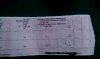
Anyone else notice the variation in T wave between the first and second beats in v4-6? The second beat looks more concave than the first
") I'd agree with the possibility hyperK+, but the QRS is still rather narrow, but I'm guessing this is a newer onset.
I'd agree with the possibility hyperK+, but the QRS is still rather narrow, but I'm guessing this is a newer onset. Anyone else notice the variation in T wave between the first and second beats in v4-6? The second beat looks more concave than the first
Depends on patient presentation, but I'd be strongly suspicious of dismissing it as "just" LVH. Something in this seems to be more of a lateral slowing, which could be ischemia. I'd transport him to a level-one cardiac center and get that ECG transmitted to the cardiologist if at all possible from the field...when in doubt, call out.
Out of curiosity, how was he presenting?
With malaise, I'd suspect the worst- AMI (LCA?) or possibly metabolic derangement/electrolyte imbalance.
Yep...electrolyte, potentially?
That's a pretty crappy tracing to call that a U wave.
I don't think I'd be overly excited about this patient from a cardiac standpoint... But I'd be working to get a cleaner 12 lead on the way in to the ED.
That isn't hyper K though. Well, the pt could have hyper K, but the EKG isn't showing hyper K.
To get technical, a dialysis pt with long term poorly controlled potassium is unlikely to show EKG changes indicative of hyperkalemia unless they have an acute spike in their potassium intake/output that causes a significant elevation in serum potassium levels. When potassium levels rise slowly, the body reacts by leeching calcium from the bones and causing a rise in calcium levels to counteract the potassium. Because of this the EKG doesn't change, or the changes are subtle compared to the potassium level.