Weird EKG.... Have at it!

jroyster06

Forum Crew Member
59
1
0
Ok guys have at it!

75 yo female c/c of not feeling well x45 min.
PMx of AMIx4
A/V on demmand pacer
Med compliant, in good shape, well groomed and appears to take care of herself well. Pt husband states they were at the cardiologist today to have her pacer checked and everything was good. Pt states that her sensation of not weeling well woke her up while she was sleeping. Pt is sitting on her porch in no apparent distress, skin is warm slightly pale.

Attendent parter applies Monitor minus 4ld at this time.
BP 138/86
P68-84ish irregular
98% RA
Respirations 16 unlabored.

Pop the 4ld on and WHALA!
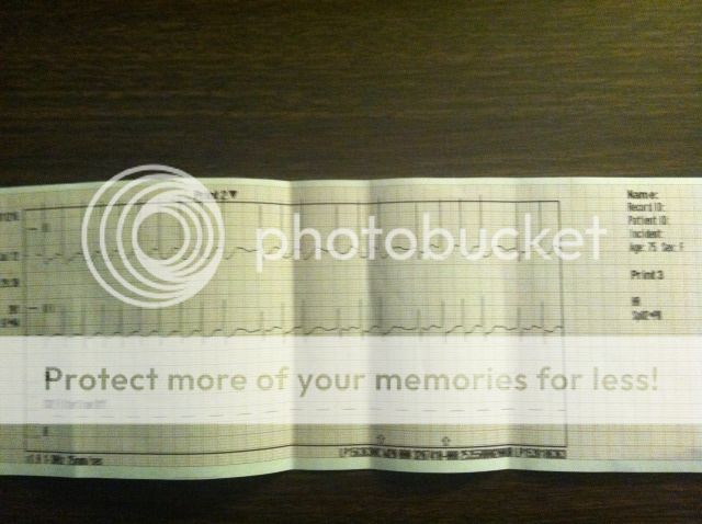
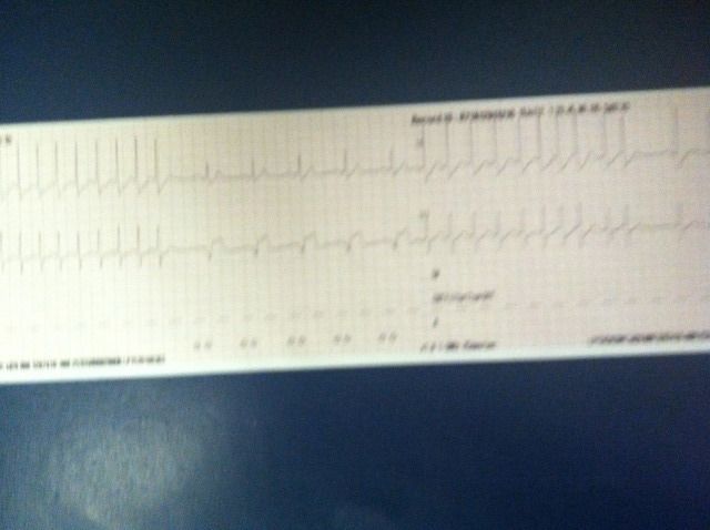
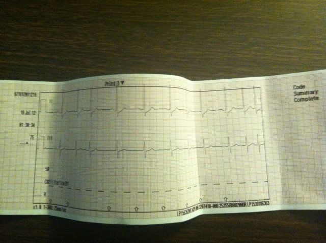
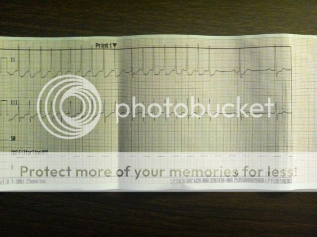
Pt heart runs in the 70's for appx 30 seconds then it runs in the 220's for about 30 seconds. And then back to the 70's....and so on. Lead placement verified.

12ld is unremarkable aside from rate.

Im sorry for the poor quality of the 4lds, It was 3am and taken with my phone. I printed a lot of strips but they were left at ER with MD. Note that when she is in a tachy rate the QRS morphology remains constant and unchanged. However, during her periods of normal rates, the QRS has several different shapes, including pacer induced complexes. I really wish i could put the whole call on here as far as the 4ld goes but i cant. What are yall thinking. My shot from the hip says aberrant conduction pathway?
IMG_0858.jpg


IMG_0856.jpg


IMG_0859.jpg


IMG_0857.jpg


IMG_0858.jpg
 
Last edited by a moderator:
OP
OP
J

jroyster06

Forum Crew Member
59
1
0
Apparently I'm not smart enough to operate Photobucket effectively enough to rotate pictures... sorry guys.
 

VFlutter

Flight Nurse
3,728
1,264
113
I can't really tell from the pictures, they are not zoomed in enough. But it looks like SR with episodes of PSVT (Probably AVRNT)

safe_image.php
 
Last edited by a moderator:

NomadicMedic

I know a guy who knows a guy.
12,097
6,845
113
I tried to march it out the best I could on the screen. It looks pretty regular to me... PSVT is my vote, a renenty .

And I'd not treat it, unless it was sustained for long periods of time...
 
Last edited by a moderator:

Medic Tim

Forum Deputy Chief
Premium Member
2,140
83
48
I tried to march it out the best I could on the screen. It looks pretty regular to me... PSVT is my vote, a renenty .

And I'd not treat it, unless it was sustained for long periods of time...

This
 

mycrofft

Still crazy but elsewhere
11,322
48
48
How'd the pt look?
 

stemi

Forum Crew Member
88
0
6
I'm thinking rapid A-fib, cause of irregularity, with weird parts of sinus rhythm in between.
 
OP
OP
J

jroyster06

Forum Crew Member
59
1
0
My first thought was afib RVR but it wasnt. Way too regular. Sorry for the poor quality. I didnt treat it other than bilat large bore's incase things got bad and transport. Pt looked in fair condition as stated, pale skin. PSVT is what i called it i just never would have thought i would see it reoccur so many times with NSR in between.
 

Doczilla

Forum Captain
393
65
28
There are no fibrillatory waves between the QRS complexes. Not A fib. When the bouts occur , its a narrow complex with t waves obscured by the next QRS complex.

When looking at a narrow complex arrythmia, any time the relationship or distance between the QRS and the P and T waves are either inappropriate or obscured entirely, PSVT is the first guess.
 

Christopher

Forum Deputy Chief
1,344
74
48
Ok guys have at it!

A 12-Lead during the tachycardia would be wonderful to help, but given the 3-leads the following are all rhythms of interest (in order by likelihood):
  • AVNRT
  • Atrial Fibrillation
  • Atrial Tachycardia
  • Junctional Tachycardia

I would stress that although the rhythm is relatively regular and the TP segment appears nice and level, perhaps this patient is in persistent atrial fibrillation with a demand pacemaker and the leads we're viewing just aren't capturing the F-waves well. Basically, I wouldn't rule out A-fib just yet.

As far as prehospital treatment, it would most likely just be supportive. If the tachycardia enjoys a sustained run an adenosine bolus would be quite illustrative, either terminating the arrhythmia outright or allowing the underlying automatic rhythm to show its face better.
 

18G

Paramedic
1,368
12
38
My vote is PSVT.

Perhaps it's possible, but for Afib to start and stop like that doesn't seem likely from my experiences anyway. And it looks more like SVT.

The patient sounded pretty stable so like Christopher said, supportive treatment only or a trial of adenosine (maybe).

Interesting patient.
 

Doczilla

Forum Captain
393
65
28
Did anyone agree with the no fibrillatory waves? The bit that might look like them in one spot looks like artifact.
 

Christopher

Forum Deputy Chief
1,344
74
48
Did anyone agree with the no fibrillatory waves? The bit that might look like them in one spot looks like artifact.

I don't disagree that the TP segment appears isoelectric, but in monitor mode in the limb leads this doesn't exclude fibrillatory waves. The R-R intervals are a bit too irregular for me to wave-off atrial fibrillation entirely. I do believe it is the less likely option.
 

Doczilla

Forum Captain
393
65
28
Alright, finally home, and could see it on a big screen.

This is for my own enrichment, cause I sure am rusty:

Dosen't the presence of monomorphic P waves also point away from a-fib?

Also, dosen't the presence of a P wave, or at least the PR relationship rule out junctional tachycardia?
 

VFlutter

Flight Nurse
3,728
1,264
113
If you take a look at the 3rd picture, the "Normal one" it appears to be SR but there is a variation of the R-R on like the 3rd complex, presumably a PAC. If you look at the last few complexes you see a normal sinus beat, then a PAC and then the start of the abnormal rhythm. So to me that leans towards the diagnosis of a re-entry rhythm like PSVT over A fib. Also if you look at how the rhythm terminates, it appears to end with another premature beat. I still think if you blow up the bottom lead you will see P waves buried in QRS-T, notice the slight variations in the QRS/ST morphology during the abnormal runs?

Could you possibly scan the images instead of taking pictures next time? Might make things clearer


Also, just to comment. A fib can definitely start and stop like this, most patients will have many frequent brief conversions back and forth such as this before they actually sustain.
 
Last edited by a moderator:
OP
OP
J

jroyster06

Forum Crew Member
59
1
0
All good thoughts.... I was thinking PSVT during transport, just what surprised me was the bouncing back and fourth. It was very interesting. ER doc said if she sustained the tachy rhythm they would start her on cardizem. Im thinking it could still be an alternate conduction pathway and was thinking an ablation was in her near future.
 

sweetpete

Forum Lieutenant
144
3
18
My .02---I'm leaning towards PSVT to a sinus rhythm. Hard to really tell, but really interesting nonetheless.

Thanks for posting!!
 

zzyzx

Forum Captain
428
90
28
Looking only at the second to last strip, I see a tachycardia going into a sinus bradycardia, so my first thought is tachycardia-bradycardia syndrome (a variation of sick-sinus syndrome).
 
Last edited by a moderator:
Top