ECG Case

Sublime

LP, RN
264
6
18


This is a 79 y/o female who was found by her daughter lying in bed difficult to arouse. She has a hx. of dementia, stroke, hypertension, and hyperlipidemia. Daughter states she is more confused than normal. Unsure of onset as she just came by her moms house and found her in bed.

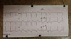
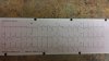
On arrival lying supine in bed. She is pale and diaphoretic. Vomits multiple times during exam. Generalized weakness present, she can't hold herself up. She is oriented to her name only. Will only answer questions occasionally but will state that she has chest pain and clutches the right side of her chest. Patient will not give a answer for the 1-10 pain scale and just moans. She appears mildly short of breath. Fine crackles heard in anterior bases. Negative stroke scale. Pulses present and regular. Sinus tach. on monitor. 12-lead shown above with posterior / right sided view on one of them.

Vitals: 156/84, 116, RR 20, 88% on room air (up to 91% on 4L via nasal, up to 97% on NRB). In this scenario the closest cath lab is 45 minutes away. A community ER is 30 minutes away. Helicopter is available. What is you decision and interpretation of ECG?
 

Attachments

  • 20140131_185521.jpg
    20140131_185521.jpg
    35.5 KB · Views: 243
  • 20140131_185436.jpg
    20140131_185436.jpg
    32.6 KB · Views: 233
Last edited by a moderator:

Anjel

Forum Angel
4,548
302
83
Pics not working.
 

MonkeyArrow

Forum Asst. Chief
828
261
63

Anjel

Forum Angel
4,548
302
83
Hmmm. I'm on tapatalk. Maybe that's why.
 

STXmedic

Forum Burnout
Premium Member
5,018
1,356
113
LMCA occlusion. She's going to the cath lab posthaste.
 
OP
OP
Sublime

Sublime

LP, RN
264
6
18
LMCA occlusion. She's going to the cath lab posthaste.

My thought as well. My interpretation was widespread ST-depression with elevation in AVR and some slight elevation in V1 & 2. I was concerned for possible LMCA occlusion or subendocardial ischemia and flew this patient to a cath lab facility.

And the pics are working fine for me on tapatalk (galaxy s4)
 
Last edited by a moderator:

medicsb

Forum Asst. Chief
818
86
28
To be honest, the story doesn't sound cardiac (AMS without hypotension or severe hypoxia) and 12-leads don't really help identify a cause for the overall clinical picture. Though STE in aVR may be considered a STEMI equivilent, but I'm not so sure in this case. Could the patient have triple vessel disease or LCA insufficiency? Absolutely, and that may be what is cause the ECG changes, and sure the patient may need to be cathed or CABG'd at some point. But, what is causing the ECG changes wouldn't really account for the change in mentation.

I'm thinking that for this patient, the 12 lead could be a red herring.

A quick quote from Dr. Smith's ECG Blog (http://hqmeded-ecg.blogspot.com/search/label/aVR):

"1) diffuse subendocardial ischemia is more likely to have non-ACS causes than traditional STEMI ECGs [they are frequently caused by stress cardiomyopathy (usually due to small vessel vasocontriction due to catecholamines) and also by demand ischemia] and

2) when due to ACS, STE in aVR is very infrequently due to coronary occlusion (there is a mistaken belief that ST elevation in aVR with diffuse ST depression is associated with left main occlusion and this is not true!). Rather it is due to coronary insufficiency due to a tight left main or 3-vessel disease with inadequate coronary flow."

So, through the retrospectoscope, I'd say that the flight was not needed (though didn't cause harm) and that it is likely that the patient could have been adequately managed at the community hospital (though, I'd not fault anyone for going the extra 15 minutes to the hospital with more comprehensive cardiac care).
 
OP
OP
Sublime

Sublime

LP, RN
264
6
18
medicsb, I appreciate the post and your perspective. I had not read the quote from Dr. Smith before, that is good information although it conflicts with some other sources I learn from.

What about the other symptoms? Do you think she might have a viral infection causing the nausea and increased hr which lead to demand ischemia leading to the ecg changes and symptoms of MI?
 

MonkeyArrow

Forum Asst. Chief
828
261
63
medicsb, I appreciate the post and your perspective. I had not read the quote from Dr. Smith before, that is good information although it conflicts with some other sources I learn from.

What about the other symptoms? Do you think she might have a viral infection causing the nausea and increased hr which lead to demand ischemia leading to the ecg changes and symptoms of MI?

Although I suppose that chain of events is plausible, it is not very likely at all. The only problem I see is with a viral infection, you would probably see an abnormal temp. The symptoms could cause increased demand on the heart and raise HR but that would theoretically only create a sinus tach assuming no underlying problems. The electrical pathology change in the ECG would be indicative more of an insufficiency problem or a MI, not a viral infection that could act that fast to create that much ST depression. If the viral infection was that bad, one would think that the pt. would go to the ED or be seen by their PCP for said viral Sx.
 
OP
OP
Sublime

Sublime

LP, RN
264
6
18
I was just trying to think of a scenario that could give her that presentation and ecg changes besides a coronary event. I agree there are more likely differentials.

I still think LMCA is highly probable though given her presentation.

Chest pain, nausea, vomiting, palor and diaphoresis coupled with gross ST-depression and elevation in AVR just point me in that direction. I would appreciate other opinions and viewpoints though.

So far I contacted the air crew and was told the facility decided to manage her medically for the time being. That's about all I got. Will try to get further information.
 

MMiz

I put the M in EMTLife
Community Leader
5,519
401
83
Sublime,

If it's okay with you, I attached the images directly to the thread so mobile members may view the files.
 

medicsb

Forum Asst. Chief
818
86
28
medicsb, I appreciate the post and your perspective. I had not read the quote from Dr. Smith before, that is good information although it conflicts with some other sources I learn from.

What about the other symptoms? Do you think she might have a viral infection causing the nausea and increased hr which lead to demand ischemia leading to the ecg changes and symptoms of MI?

There is a lot of things to consider. Infectious or metabolic etiology would be high on my differential. SSx of MI overlap with many many other disease processes. CP itself has a pretty long list of causes - costochondritis, aneurysm, pneumonia, PE, esophageal spasm, GERD, pneumothorax, etc. N/V can be caused by head injury, infection, CNS issues, pain, toxins, GI disease, etc. This is why patients can require such exhaustive work-ups, especially when they're altered.

But, yeah, it could have been demand ischemia. She certainly has risk factors for CAD. The ECG changes also could have been old. It's possible that she could have a known left main coronary disease and that surgery or intervention was decided against due to dementia.
 

MSDeltaFlt

RRT/NRP
1,422
35
48
My impression is she's septic and not cardiac. AMS on a geriatric with PMH of stroke and dementia who is pale, diphoretic, vomits, and has rales with mild hypoxia? And she goes to a cath lab?!? That's sepsis until proven otherwise.

The ECG looks old to me. Sinur rhythm with abbarancy with an ectopic beat. No STEMI. No axis deviation. Chronic changes only. What was her temp?
 
Last edited by a moderator:

medicsb

Forum Asst. Chief
818
86
28
I was just trying to think of a scenario that could give her that presentation and ecg changes besides a coronary event. I agree there are more likely differentials.

I still think LMCA is highly probable though given her presentation.

Chest pain, nausea, vomiting, palor and diaphoresis coupled with gross ST-depression and elevation in AVR just point me in that direction. I would appreciate other opinions and viewpoints though.

So far I contacted the air crew and was told the facility decided to manage her medically for the time being. That's about all I got. Will try to get further information.

You're anchoring on the ECG and conforming a portion of her SSx into an ACS diagnosis, while ignoring the change in mental status (which is essentially the chief complaint). Only in certain circumstances are you going to get a change in mental status with ACS/MI - drop in cardiac output leading to decreased perfusion of the brain or hypoxia from pulmonary edema (I would doubt that 88% in and of itself would produce much of a AMS). Given the patients age and dementia history, it is possible that the moderate hypoxia could have caused the patient to develop delerium, so in that way a primary cardiac event could have produced AMS. However, it's not something you want to get caught on considering the number of life threats that are not ACS that could produce her constellation of symptoms (big one being sepsis from pneumonia, which could account for her CP).

And as said before you're probably right that the patient has LMCA disease, but that is not likely the root cause of the patients problem when you met her.

And if the root cause was ACS/MI, it would take some time to determine that, and then considering her dementia, it would need to be discussed with family whether or not you want to rush her to the cath lab for intervention. Also, given the unknown time of onset, it is possible PCI may not be helpful at all, and would certainly risk worsening her mental condition.

Anyhow, as a medic, this was a difficult case. There is no slam-dunk answer, at least not in the prehospital phase.
 

VFlutter

Flight Nurse
3,728
1,264
113
Not sure about your area hospitals but this patient would probably not get an emergent cath here. They would be sent to tele/ICU and maybe go to the CCL the next day unless there was a large troponin bump or the schedule was light.

Just curious, Why a NRB on a patient who is AMS and actively vomiting? Seems like a high risk for aspiration (which may be the cause of the possible pneumonia). 91% on 4L NC, I would have throw it up to 6L and let it ride.
 
Last edited by a moderator:

MSDeltaFlt

RRT/NRP
1,422
35
48
A few more questions to ensure we are treating the patient and not some cotton-pickin' monitor.

1. What was her BGL?

2. EtCO2?

3. Chest pain tender to palpation?

4. Foul smelling urine? Remember. Geriatric FEMALE with dementia. Always rule out UTI.

5. Skin turgor?

6. Oral mucosa moist or dry?

This scenario presentation is lacking several serious clinical assessment findings.
 
Last edited by a moderator:
OP
OP
Sublime

Sublime

LP, RN
264
6
18
My impression is she's septic and not cardiac. AMS on a geriatric with PMH of stroke and dementia who is pale, diphoretic, vomits, and has rales with mild hypoxia? And she goes to a cath lab?!? That's sepsis until proven otherwise.

The ECG looks old to me. Sinur rhythm with abbarancy with an ectopic beat. No STEMI. No axis deviation. Chronic changes only. What was her temp?

I think sepsis is a good differential, and in retrospect I should have considered it. Not going to lie I probably tunnel visioned on the ECG and ACS route. I can't say I agree with your interpretation of the strip though. After thinking about it and hearing interpretations from others, I think it is very plausible this patient had severe underlying CAD such as multi-vessel disease or left main stenosis that was exacerbated secondary to an underlying condition such as sepsis resulting in ischemic changes. But to just call it "chronic changes" and write it off as sinus rhythm with abberancy I think is discrediting the diagnostic value of the ECG in this case.

You're anchoring on the ECG and conforming a portion of her SSx into an ACS diagnosis, while ignoring the change in mental status (which is essentially the chief complaint). Only in certain circumstances are you going to get a change in mental status with ACS/MI - drop in cardiac output leading to decreased perfusion of the brain or hypoxia from pulmonary edema (I would doubt that 88% in and of itself would produce much of a AMS). Given the patients age and dementia history, it is possible that the moderate hypoxia could have caused the patient to develop delerium, so in that way a primary cardiac event could have produced AMS. However, it's not something you want to get caught on considering the number of life threats that are not ACS that could produce her constellation of symptoms (big one being sepsis from pneumonia, which could account for her CP).

And as said before you're probably right that the patient has LMCA disease, but that is not likely the root cause of the patients problem when you met her.

And if the root cause was ACS/MI, it would take some time to determine that, and then considering her dementia, it would need to be discussed with family whether or not you want to rush her to the cath lab for intervention. Also, given the unknown time of onset, it is possible PCI may not be helpful at all, and would certainly risk worsening her mental condition.

Anyhow, as a medic, this was a difficult case. There is no slam-dunk answer, at least not in the prehospital phase.

Once again, thanks for your input. I contributed the worsening mental status to the hypoxia at the time.

Not sure about your area hospitals but this patient would probably not get an emergent cath here. They would be sent to tele/ICU and maybe go to the CCL the next day unless there was a large troponin bump or the schedule was light.

Just curious, Why a NRB on a patient who is AMS and actively vomiting? Seems like a high risk for aspiration (which may be the cause of the possible pneumonia). 91% on 4L NC, I would have throw it up to 6L and let it ride.

Non-rebreather was placed after an IV was established, Zofran was given, and the patient was no longer actively vomiting.

A few more questions to ensure we are treating the patient and not some cotton-pickin' monitor.

1. What was her BGL?

2. EtCO2?

3. Chest pain tender to palpation?

4. Foul smelling urine? Remember. Geriatric FEMALE with dementia. Always rule out UTI.

5. Skin turgor?

6. Oral mucosa moist or dry?

This scenario presentation is lacking several serious clinical assessment findings.

1. 140 something
2. Did not obtain
3. Not tender to palpation that I could tell (her moaning did not change).
4. No urine noted. Did not ask daughter about it.
5. Normal for an 80 y/o
6. Moist
 
Last edited by a moderator:

VFlutter

Flight Nurse
3,728
1,264
113
I am favoring Demand Ischemia with underlying CAD as opposed to a primary coronary event.
 

MSDeltaFlt

RRT/NRP
1,422
35
48
Here's why I call the ECG chronic. There is no axis deviation. No LBBB or RBBB. Though there is ST elevation in V1, it is only in V1 and even then it is variable. No ST elevation in V4R. And variable ST depression in V8 & V9. Yes, this little old lady may have multi-vessel disease. But I wager she has been having said multi-vessel disease for quite some time. Er go chronic changes. She's septic.
 
Last edited by a moderator:
Top