12 Lead EKG interpretations: let's do it!!!

clibb

Forum Captain
366
1
0
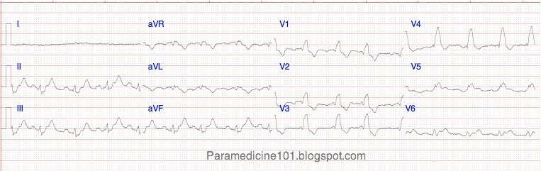
clibb, I would agree that it looks like a first degree block, or possibly even a wenckebach(hard to tell without seeing a couple more seconds) after the patient converted.

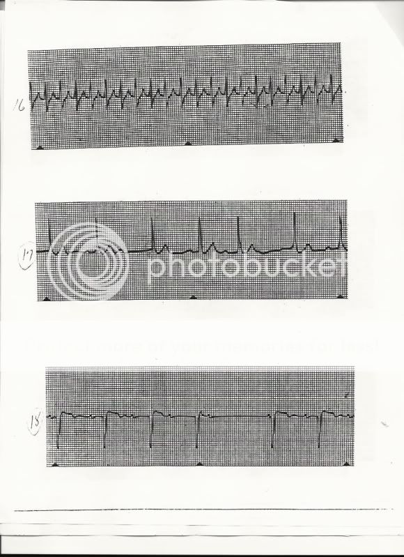
If you look at lead II.
It goes from V-Tach to Cuplet PVCs. Then the T waves aren't consistent at all. ST segment looks normal or I would think MI.
If you go to the fourth T-P waves after the V-tach on Lead II the T and P waves and how it the line declines is something I'd be worried about.

And then it looks like it leads into the heart blocks.

On lead I and lead III, I would totally agree with the Wenckebach.


These are fun. I have like 100 strips from my EKG class if you guys want me to post them.
 

clibb

Forum Captain
366
1
0
Here you go. Three different strips. Might be a little too basic, but still fun.
Let me know if you need a more "zoomed in" picture.

Strip1-3.jpg
 
OP
OP
Dutch-EMT

Dutch-EMT

BScN
97
1
8
I see a BBB (clear in V4 and V5), so that makes it not possible to diagnose on MI based on ST.
There are ST elevations in the precordial leads, but with a BBB not reliable for diagnose based on this ECG.
 

FL_Medic

Forum Probie
10
0
0
I see a BBB (clear in V4 and V5), so that makes it not possible to diagnose on MI based on ST.
There are ST elevations in the precordial leads, but with a BBB not reliable for diagnose based on this ECG.

That is entirely incorrect, but I am not surprised to see a response like this. Well, not entirely, they both present with BBB patterns, and there is ST-Elevation. The assumption that you can not evaluate ECGs with BBBs for STEMI is incorrect.

LBBB presents with negative QRS and T-wave discordance which can mimic a STEMI. There are still ways to interpret LBBB ECGs for STEMI, search Sgarbossa's criteria. I use a simple rule though. If the STE is > 20% of the depth of the preceding S-wave, AMI is likely. Also, convex STE is usually an ominous finding, even with LBBB.

RBBB presents with positively deflected QRS complexes and T-wave discordance which does not cause elevation of the J-point/ST-segment. The problem some have with RBBB is that they are not evaluating the true width of the QRS complex due to funky looking morphology. This is interpreter error however, and does not mean you can't interpret a RBBB ECG for STEMI.
 
OP
OP
Dutch-EMT

Dutch-EMT

BScN
97
1
8
I agree with diagnose MI when there is RBBB.
But when there is LBBB and you suspect a MI, will you start treatment with meds? An echo and/or lab (CK and troponine) will confirm MI.
 

FL_Medic

Forum Probie
10
0
0
I agree with diagnose MI when there is RBBB.
But when there is LBBB and you suspect a MI, will you start treatment with meds? An echo and/or lab (CK and troponine) will confirm MI.

Well think about the treatments we do for MI. Transport to a PCI capable facility, ASA, & NTG. A little morphine too. Who will this hurt? Some, but not most.

So my answer is yes.

Concluding that you have changes indicative of a LBBB with STEMI should be no different when considering treatments, from any other STEMI.

From our friend Tom B. <<-- go read, you won't regret it.

Also, check out this link to better diagnose BBB/IVCD. I believe looking at V4 & V5 for bunny ears is not beneficial.
 
Last edited by a moderator:
OP
OP
Dutch-EMT

Dutch-EMT

BScN
97
1
8
Well think about the treatments we do for MI. Transport to a PCI capable facility, ASA, & NTG. A little morphine too. Who will this hurt? Some, but not most.

So my answer is yes.

Concluding that you have changes indicative of a LBBB with STEMI should be no different when considering treatments, from any other STEMI.

From our friend Tom B. <<-- go read, you won't regret it.

Also, check out this link to better diagnose BBB/IVCD. I believe looking at V4 & V5 for bunny ears is not beneficial.

Interesting link!!!
 
OP
OP
Dutch-EMT

Dutch-EMT

BScN
97
1
8
Time for a new ECG?

I copied this one yesterday.

The story behind this ECG:
A lady, 90 yrs old.
30-5-2010 she felt down and ended with a pertrochanter femurfracture.
She had surgery the same evening.

The 1st of june early morning her heartrate was goïng faster.
NIBP between 95/55 and 125/60, fluctuating the whole morning.
An ECG was made. Temp: 37,0. Also lab (hb/ht/KNUK/ery's/leuco's/CRP) was token.

36309005.jpg


For the large version click HERE
 

clibb

Forum Captain
366
1
0
Dutch EMT,

When was her previous MI? Or is that the aftermath of the heart surgery I'm seeing with the declined ST segment?
Obvious 1st degree heart block. I'd say her heart rate is between 180-220 so I'd say her rhythm is SVT.
 
OP
OP
Dutch-EMT

Dutch-EMT

BScN
97
1
8
Patiënts History:
-Only a bad spine caused by age and operated by a plastic surgeon (triggerfingers)
-no history at all, no MI in the past.

Patient looks a bit tired but is alert. No cheastpain or other complains...
 

Shishkabob

Forum Chief
8,264
32
48
Here's one for ya---- 73yo female, no history of heart disease.

First image is a 3lead, second 2 images are the 12lead.
20100606130355.jpg


83300214.jpg


33082114.jpg
 

TomB

Forum Captain
393
82
28
Sinus bradycardia with 1AVB and nonspecific intraventricular conduction defect (LBBB morphology in lead V1 but S-wave present in lead I). QRS duration is at least 160 ms, approaching 200 ms. Consider hyperkalemia. What is the chief complaint, history, physical exam, etc.?

Tom
 
OP
OP
Dutch-EMT

Dutch-EMT

BScN
97
1
8
Sinusbradycardia, about 50 bpm. 1st degree AV (>200msec)
Axis: I isoelectric, aCF positive, so heartaxis is normal, but about 90degree and heading to the right...

R-R1 in II (small), III, avF and a BBB pattern in V1 and V2.

I see a wide S in I and III in the 3 lead. Also wide QRS. Low R progression.

Can it be some kind of combination of BBB, 1st dgr AVblock and fascicular block.
QRS wider and low R fits hyperkalemia, but I don't see a Higher and sharper T.
 

Shishkabob

Forum Chief
8,264
32
48
Sinus bradycardia with 1AVB and nonspecific intraventricular conduction defect (LBBB morphology in lead V1 but S-wave present in lead I). QRS duration is at least 160 ms, approaching 200 ms. Consider hyperkalemia.
Sweet... for my first true emergent 12-lead alone in the field, I was right! I feel happy now :D

What is the chief complaint, history, physical exam, etc.?

Tom


CC was of a 70s yo female post fall in room, going from lethargic to completely unresponsive (GCS 3) and back again.


Pt had no prior cardiac history of arrhythmias, PAD, CAD or HTN. No hx of CVA either. Was hospitalized for pneumonia a while before but cleared up with Rifampin. Pt type 2 DM.

Physcal was unremarkable on everything but the diabetic sores she had on her legs.
 
Top