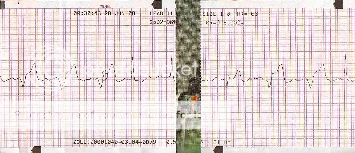
WTH at this rhythm (56K, no)

jedirye

Forum Lieutenant
Messages
114
Reaction score
0
Points
0
So I'm on a clinical and around midnight or so we get called for an OD. Guy is a 79 Y/W/M pt who intentionally ingested an unknown amount of hydrocodone (prescription x 2 years ago) to intentionally hurt himself. Pt is found sitting by toilet dry heaving, nothing in bowl (no pill fragments, etc.) Pt is A/Ox4 with a GCS of 15. Pt ingested the unknown amount (bottle stated 20 pills) over the course of three hours. Get him in the truck, hook him up and I see that crazy rhythm. Hx of patient? Pacemaker, yes, but I see no pacer spikes on the strip. Pt also had an ablation done for I believe tachycardia. Pt denies any cheif complaint at the time other than N/V (no chest pain, SOB, etc.) All complexes are perfusing, by the way. I'm riding with a seasoned vet. who looks at the screen and says no big deal. I insist on doing a 12-lead regardless and the one included was the best I could get (and that was with the truck stopped!). I brought this strip into work with me to see what other medics had to say because I am still baffled by it. No one has given me a definitive answer. I brought the strip with me to the next clinical and asked the seasoned vet. what he thought it was as well. He gave me some round-a-bout answer and didn't really give me a definitive answer either. Any takers??

Also, pardon the writing, the seasoned vet marked my strips! :glare:


Scan.jpg


Scan2.jpg


Scan3.jpg


Scan4.jpg


Appreciate any insight and help. I'm still learning here (and forever will)...


-rye
 
This patient has serious conduction problems.. It appears that there is complete AV disassociation or 3rd degree block. The wide complex QRS beats are paced beats. The pacing spike is very hard to see with today's demand pacemakers. The narrow complex QRS's are the patient's native escape beats, and are junctional. That's my story and I'm sticking with it ;)
 
looks like A-fib with preexisting left BBB. i get more to you when i get off work.
 
I thought Afib too.. but the rhythm is totally regular... thus.. not afib.. and there are regular P waves visible in some of the leads....
 
i just can't be sure that they are p waves due to their morphologies.
 
there is a lot of artifact to sort thru all right... the other thing is, I'm not aware of a bundle branch block being intermittant with a regular rhythm. There are all kinds of rate dependent arrhythmias and ectopies, but in this case, with the regular rate, I can't see a bbb in most beats with narrow QRS beats inbetween. The wide QRS is the same each time and is very typical of ventricular demand pacemaker beats. I can't see a V5 or V6 - often pacemaker spikes are most apparent in those - don't know why. There - talked myself into it.. ;-0
 
Last edited by a moderator:
To be honest, I was thinking third degree also but when I suggested that I was basically laughed at due to the rate............

Of coure they thought it was Second degree II, even though there are varying PRI's............... Still taking suggestions I guess.

-rye
 
2nd degree type II has the same P-R - so you were right... they are wrong. (I'm a humble type huh?)
 
I would meander to say that a consult with the cardiologist would be in order. My first question when presented with a strip is always: "was the patient symptomatic (i.e. Decreased LOC/GCS, Abnormal vitals and decreased cerebral perfusion, etc.) If the reply is negative, then my answer is treatment is supportive (O2, monitor, IV, mobile to hospital)

I see you pack a Zoll (and a picture of a LP in one of the strips??). I would agree with some of what has been said so far. Pacemaker spikes is not always apparent, is depends on whether the pacemaker is actively performing its duty at the time of interpretation or recording (do yourself a favour and check up on the different type of pacemakers, and also check to see if the patient knows which one he has, they normally don't).

Although the rhythm is irregular, it can also be classed as being regularly irregular (Regular periods of irregularity). As mentioned, this is way too regular (at times) for being an A-Fib, hence that one is out. Looking at the origan of the beats (its good you compared the electrical representation of the monitor with actual beats providing output, i.e., does the pulse correspond with the monitor?) the majority of them are widened, which implies ventricular in nature.

Now, the escape beats... I am not convinced the narrow complex beats are the escape beats (I could be wrong, and I will consult some more texts). Should the conduction not follow through the normal pathway, or be blocked, it would be sensible to say that the ventricles would generate some form of electrical activity (as any cardiac cell has the ability to do so), in order to try and maintain some form of cardiac output, hence those rather being the escape beats. I could not find any reference to escape beats in the text I had at hand, but it would be wise to check on the measurement of the QTc interval, as that is what determines "Life saving" escape beats from non-lifesaving escape beats.

The 12 lead (?performed by EMS) yip, there is a lot of interference, and as per the previous strips, LII is the only to really use, in order to go into the nitty grities. It would have been interesting to see the the in hospital 12 leads machine had to diagnose this as!! LBBB, I can see the resemblance, the "bunny ears" however BBBs are typically diagnosed in V1 and V6 (?v1 WiLLiaM v6:v1 MaRRoW v6?), and there is not a lot evidence in these leads, as well as you have to take the interference into consideration. If I had to put a BBB to the diagnosis, it would be Left though.

Diagnose this?? I would also go with the 3rd deg AV block. Treatment, ...Fixed transcutaneous pacing , only should the patient become unstable, otherwise supportive. Excuse the spelling Kaisu.
 
I see you pack a Zoll (and a picture of a LP in one of the strips??). I would agree with some of what has been said so far.

I had a feeling someone was going to comment on that. Actually, I pack LP12's, but who I am doing my ride-a-longs with pack Zoll's. The LP in the background is my textbook I used to hold the strips down.

-rye
 
They kind of look like ventricular capture beats. still the p-r interval has too many irregularities and morphologies. p waves are still looking morphed to me.
 
No one else?
 
I would go with 2nd Type II with either runs of idoventricular beats or runs of abbarent conduction. The 12 Lead really makes it difficult, but looking at the way the complexes go on the V leads, I'm leaning toward runs of idioventricular beats.
 
But how in the world can it be Second Degree II given the PR intervals??

-rye
 
If you notice the intrinsic rate drops down when you have the fusion beats occurring, that is why it is coming from multiple foci. The wide bizarre complexes (fusion/ventricular escape beats) is attempting and does successfully convert it back to a S.R.

Too much BS in the baseline to determine other factors and as the say "non-contributory" and insignificant.

Although, I do doubt the scenario as mentioned caused the arrhythmia, it should raise flags for other possibility such as tricyclic antidepressants, etc.. which can cause such arrhythmias.

R/r 911
 
I meant 2nd Type I. Sorry. Typo.
 
Back
Top