Reciprocal Changes in 12-Lead with Acute STEMI

Jayy

Forum Probie
Messages
21
Reaction score
1
Points
3
I'm trying to understand reciprocal changes leading to the interpretation of a Posterior wall MI.

Is it basically if you have ST elevation in the inferior leads + reciprocal depression in the Anterioseptal leads, that automatically means it's a posterior wall MI? This is a concept we are just being introduced to in school, so I apologize if I'm not using proper terminology.

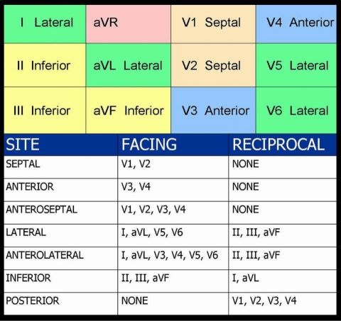
Here's the guide we were given to review:

stemi.JPG
 
The inferior leads (II, III and aVF) have nothing to do with the posterior. The thing that makes this EKG suspicious for posterior MI is the ST depression in v1-v3. If you put posterior leads on (v7-v9), you'd likely see ST elevation there. Leads v1-v3 are reciprocal to the posterior leads.

There also happens to be an inferior MI going on as you can see from the limb leads.

Think about the limb leads and precordial leads as looking at different planes of the heart: I, II, and III (and aVR, aVL and aVF which are derived from I, II, and III through some math) look at the heart vertically, while the v leads look horizontally. They don't really have anything to do with each other.

This picture might help. Notice how the v leads circle around the heart horizontally while the limb leads go around vertically:

ECG-Anatomy-LITFL.jpg
 
I'm trying to understand reciprocal changes leading to the interpretation of a Posterior wall MI.

Is it basically if you have ST elevation in the inferior leads + reciprocal depression in the Anterioseptal leads, that automatically means it's a posterior wall MI? This is a concept we are just being introduced to in school, so I apologize if I'm not using proper terminology.

Here's the guide we were given to review:

View attachment 2112

If the only abnormality on this ECG was the abnormality present in leads V1-V3 I would call it acute isolated posterior STEMI.

It is true that you can "train your mind" to see acute isolated posterior STEMI by paying close attention to these leads in the setting of acute inferior STEMI because the posterior descending artery often branches off the RCA.

Tom
 
@Gurby: A couple cautions. First, there is indeed a relationship between the posterior and inferior leads; they are often largely fed by the same vessel. Second, it is something of an overstatement to suggest that reciprocal leads are unrelated anatomically; most of them can be associated if you recognize which walls actually face one another.
 
@Gurby: A couple cautions. First, there is indeed a relationship between the posterior and inferior leads; they are often largely fed by the same vessel. Second, it is something of an overstatement to suggest that reciprocal leads are unrelated anatomically; most of them can be associated if you recognize which walls actually face one another.

Good point about vessels - that's something I need to study more. Like how whenever you have an inferior MI you're also suspicious for RV MI because it could also be affected depending on where the RCA got blocked off. I guess it's a big overstatement to say that "they don't have anything to do with each other", but I think for the OP's question, for someone just starting to learn EKG's, it's okay to think about them separately?

My thought process was basically this: "there is STE in II, III, aVF, with reciprocal changes in I and aVL? Okay, there's an inferior STEMI" --- "There is ST depression in v1-v3? Suspicious for posterior involvement, and because of presence of inferior MI we are extra suspicious" --- "STE in III is higher than II? This guy may also have RV involvement."
 
Sort of, yeah. My point is that certain anatomical regions "going together" is just like certain leads within one region "going together"; it's an important aspect of recognizing a pattern consistent with acute injury. You'd be missing the big picture if you said something like "well, there's elevation in V1, V2, and V3," but didn't recognize that those represent contiguous leads of the same anatomical region (the anterior wall). And likewise you're missing something if you say "there's inferior elevation; and on an unrelated note there's significant anterior depression too." It's not particularly important to be able to recognize which aspects of the RCA are occluded, but it IS important insofar as it adds supportive data to your diagnostic constellation (RCA occlusion), and -- particularly in more subtle cases -- that is always a good thing.

I do remember being taught early that the anterior wall is reciprocal to the inferior wall and finding this confusing, because it's not especially true. (It is poorly reciprocal if anything.) The lateral wall, meaning aVL and I, are the true reciprocals for inferior injury. This type of big anterior depression is usually from posterior involvement.
 
3af11064ab360c01ba20d3d4555c6655.jpg


this chart might help as well, and provide a good cheat sheet
 
sometimes you will st elevation in the lateral lead along with the inferior lead with deep st depression in v1-v4 if you see this simply put extra leads to the back known as v7 - v9, following it around until your v9 lead in just below the left scapula.
 
Back
Top