Arrhythmias in pt's receiving blood

OKparamurse

Murse 'n medic
Messages
63
Reaction score
2
Points
8
So I had a first last night, I had a mid-50s male pt who had a GI bleed and was being transferred to a larger facility approx. an hour and a half away. Long story short, he had 500cc of blood hanging infusing at a rate of 150cc/hr. From report from the nurses, the blood had been infusing for about 20 minutes prior to the transfer. Well I put him on the monitor and I get one of the absolute weirdest tracings I've ever seen. I could only describe it was an irregular polymorphic v-tach. He would go into runs of it for maybe 5 or 6 seconds, return to his baseline (A-fib RVR @ 120s), and then go into another run roughly 20 seconds later. This continued for probably 30 minutes, and then he remained in A-fib with no PVCs or unusual runs for the rest of the trip. No hx of such from sending facility. No complaints of pain or discomfort during that time. Anyone else seen anything like this before?
 
Was this his first unit of blood or did he receive multiple prior? Not really any reason he would have an arrhythmia solely because of the transfusion but patients can become profoundly hypocalcemic after multiple units of blood. Could be coincidental or could be his heart being irritable from the anemia.
 
First unit. A little on the road research at the revealed about (If I remember correctly) 1-2% of pts going into arrhythmias due to blood transfusions, but it seemed to be either Afib or Vfib, not so much this really odd rhythm I saw. Labs were all normal at least prior to transport other than low H&H.
 
It could also be runs of RVR through an aberrant pathway and not related to the blood. What product were you giving? I'm used to smaller bags for my PRBCs.
 
You said A-fib with RVR could it had been a-fib with aberrancy ? Was a 12-lead obtained ?

I like chase's response. People who normally have A-fib are prone to wierd rythems when things get out of wack ie. dehydration, low perfusion, and electrolyte problems.
 
So I had a first last night, I had a mid-50s male pt who had a GI bleed and was being transferred to a larger facility approx. an hour and a half away. Long story short, he had 500cc of blood hanging infusing at a rate of 150cc/hr. From report from the nurses, the blood had been infusing for about 20 minutes prior to the transfer. Well I put him on the monitor and I get one of the absolute weirdest tracings I've ever seen. I could only describe it was an irregular polymorphic v-tach. He would go into runs of it for maybe 5 or 6 seconds, return to his baseline (A-fib RVR @ 120s), and then go into another run roughly 20 seconds later. This continued for probably 30 minutes, and then he remained in A-fib with no PVCs or unusual runs for the rest of the trip. No hx of such from sending facility. No complaints of pain or discomfort during that time. Anyone else seen anything like this before?


A patient like this could develop atrial fibrillation for any number of reasons. As others have mentioned, electrolyte abnormalities are common with the administration of blood. This could certainly be a cause or a contributing factor.

Another mechanism of initiation of a-fib is acute atrial stretch. Even though the patient has not had a large amount of blood administered, chances are pretty good that he got some degree of resuscitation with either crystaloid or colloid. Administration of crystaloid means the heart is seeing a larger preload, and thus creating an element of acute stretch on the wall of the atria.

Very likely it's multifactoral, encompassing two or more of these etiologies.
 
Unfortunately this is all I could find from monitor archives. And yes, this is a pretty clean tracing of his rhythm after an electrode change and after skin prep x2. I had to stare at the monitor, reposition and change electrodes, and have my partner pull over so I know it wasn't bumping down the road.
 

Attachments

  • 20161026_113604.jpg
    20161026_113604.jpg
    1.6 MB · Views: 272
Unfortunately this is all I could find from monitor archives. And yes, this is a pretty clean tracing of his rhythm after an electrode change and after skin prep x2. I had to stare at the monitor, reposition and change electrodes, and have my partner pull over so I know it wasn't bumping down the road.

My money is on equipment failure after seeing that.

That's artifact, no if ands or buts about it.

Where were your leads placed at?


Sent from my iPhone using Tapatalk
 
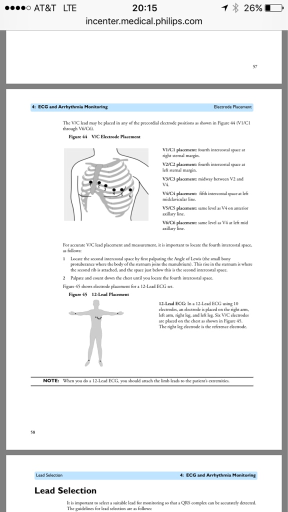
Outer pectorals and left and right lower abd. As Phillips recommends as they are "torso leads" and not limb leads. Repositioned with electrode change as well to move to thighs and shoulders. Same rhythm sitting absolutely still. And I wouldn't think machine malfunction to it just steadily clear up over time throughout transport. And by clean up, I mean these "runs" become less frequent and eventually terminate. I know you guys must think artifact or machine error, and I know this is clichè, but you just had to be there. Lol
 
Outer pectorals and left and right lower abd. As Phillips recommends as they are "torso leads" and not limb leads. Repositioned with electrode change as well to move to thighs and shoulders. Same rhythm sitting absolutely still. And I wouldn't think machine malfunction to it just steadily clear up over time throughout transport. And by clean up, I mean these "runs" become less frequent and eventually terminate. I know you guys must think artifact or machine error, and I know this is clichè, but you just had to be there. Lol

The problem is, that's not a perfusing rhythm or a rhythm compatible with life. It's not even VF. That's why I say equipment failure (notice equipment, not provider).

Philips recommends limb placement for their leads.

Source: http://incenter.medical.philips.com...re_versions_(ENG).pdf?nodeid=9929206&vernum=1

e03c3e5d77531796ebb2cdf7ac8c9389.png


Sent from my iPhone using Tapatalk
 
Ah okay. We actually had a Phillips representative do an in-service last year when we switched from LPs and he was the one that recommended the torso lead configuration. Either way, that might explain for some axis deviation, but I can't imagine it explains the grossly abnormal EKG recorded.
 
To me that also looks exactly like you've got some loose leads or some wires that are degrading to the point where intermittent contact is being made. If you're 100% sure that it's not an equipment issue, try using an alternate chest lead. One of my favorite ones to use is MCL1. With good placement, you won't get much skin movement artifact and you shouldn't pick up too much Pecs Major artifact either. If that doesn't work well, there's always the Lewis Lead... but this 100% assumes that the equipment is good.
 
To me that also looks exactly like you've got some loose leads or some wires that are degrading to the point where intermittent contact is being made. If you're 100% sure that it's not an equipment issue, try using an alternate chest lead. One of my favorite ones to use is MCL1. With good placement, you won't get much skin movement artifact and you shouldn't pick up too much Pecs Major artifact either. If that doesn't work well, there's always the Lewis Lead... but this 100% assumes that the equipment is good.

You can always try the MSL.

http://www.ems1.com/ems-products/ap...s/430502-Top-Ten-Things-Kelly-Cant-Do-in-EMS/
 
Sometimes the problem isnt where the leads meet the patient, its where the leads connect to the device. With the philips and LP where you connect the 4 lead and 12 lead can get damaged or dirt in it and create a problem.
 
Back
Top