Another 12 lead and fall scenario

Anjel

Forum Angel
Messages
4,548
Reaction score
302
Points
83
Dispatched to an independent living facility for a fall secondary to a possible heart attack at 0530.

Arrived on scene to find an 89 year old male patient laying supine on the floor of his bathroom. Pt A&Ox4 and in no apparent distress. There is a good size pooling of blood against the wall when you first walk in, that is coagulated. Blood running down the wall as well. Pt has a 2 inch laceration to the top of his head that is still bleeding, and bright red blood coming from his right ear. Unable to find laceration. Pt had dried blood all over his face as well.

Pt states the last thing he remembers was at 0130 he got up to go to the bathroom. He doesn't remember falling. The next thing he does remember was rolling over to his pull cord by the toilet and calling for help- 4 hours later. He says his defibrillator went off 2 times during the night and he thinks he is having a heart attack. He denies any chest pain or difficulty in breathing. He does have cervical tenderness. Pt is on Plavix.

Hx: Pacemaker/defib, CHF, anxiety, diabetes.

Meds: Plavix, Asa, Aldactone, xanax

So here is the 12 lead. I tried everything to get the limb leads to read better and they wouldn't. He was skin and bones so I don't know what the issue was.


avubemep.jpg


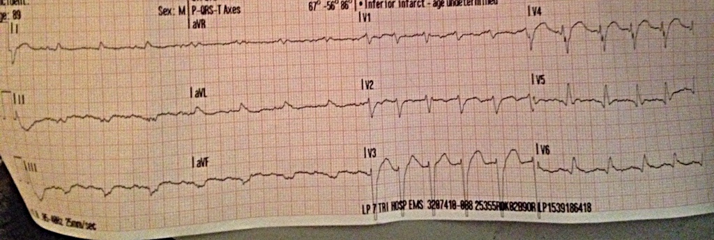
I was confused as to what was going on in V3 and V4. I don't see any pacer spikes, and my limb leads weren't very helpful.

Idk what say you guys? What would your course of treatment be?
 
So I don't know how this got into Advanced Medical but if someone could move it for me. I'd appreciate it.

@Chimpie @DEmedic
 
Moved to Scenarios.
 
Thanks!
 
I'd try another printing because that looks like crap. He'd most likely be a trauma activation. Other than that, I got nothin'.
 
I'd try another printing because that looks like crap. He'd most likely be a trauma activation. Other than that, I got nothin'.

That was like my 4th one. He was laying flat on a bathroom floor when I got it. Then again in the ambulance.

I guess later on in the ER he kept going into 10sec bouts of VTach.

Left me scratching my head.
 
You could argue for inferoposterolateral changes here with some high lateral reciprocals. Very subtle. Serial ECGs.
 
Low voltage can be due to myxedema (hypothyroidism), obesity, pleural effusion, pericardial effusion, etc. Typically <5 mm in limb leads or <10 mm in precordial leads is considered low voltage.

In the precordial leads, it looks irregular to me. No discernible p-wave in lead V1. The rate is somewhat fast. Probably atrial fibrillation.

The R-wave progression, widening of the complexes, and left axis deviation. Left anterior fascicular block.

It doesn't look like an MI to me. I wouldn't activate the cath lab for this.
 
Couple things...

1. Low voltages are common in COPD as well.

2. Other than the second beat in V1-V3, this looks regular. But I haven't marched it out so anybody who cares enough to find their calipers wins this one. I also see P waves, but I have been known to hallucinate when drinking this stuff.

3. ST changes should be interpreted by the wise in relation to their corresponding QRS. Small complexes, small changes. I would do serial tracings, although ACS is certainly not all that I'd consider with this patient.
 
The rhythm was the tough part to me.

I felt like going with irregular because the complexes almost instantaneously goes from being about 3 big boxes apart to about 2 big boxes apart. I saw that when I was trying to figure out the rate, which varies from like 90 to 137. It's regularity reminds me of atrial fibrillation with digoxin (even though that is not listed the in patient's history). At faster rates, it can be more regular looking too.

When I tried looking for p-waves, I had those moments "that kinda looks like a p-wave" and "Oh, that kinda looks like one too".Seems legit too cause what looks like p-waves sometimes appear right before the complex right about where you expected a p-wave at too. I've been bit before by atrial fibrillation though. Look at lead I. You see something you might call a p-wave twice. Where for the middle complex? The p-wave isn't obvious in lead V1 either? Overall, I felt like this rhythm was more likely atrial fibrillation than sinus rhythm.

I kinda wanna hear what Anjel's interpretation of the rhythm was when she looked at the monitor or what the machine said. Seems more interesting to me, haha! I also had a tougher time calling this atrial fibrillation because the patient isn't taking coumadin and/or digoxin (I know it's not a blood thinner, but if this is atrial fibrillation, it's pretty regular without treatment), which is what I would expect.
 
Last edited:
It was hard for me. I ended up transmitting the EKG to the hospital and going " I dunno". Because it looked like two completely different rhythms.

When monitoring in lead 2 the monitor couldn't even pick up a HR because of the low voltage. Manually it was about 92.

But when you look at the precordial leads- especially V3 and V4 it almost looks ventricular to me.

I ended up calling it a sinus rhythm in my report. Because of the regularity in the pulse and I did see p waves in my limb leads. It just all changes in the precordial.
 
So when I stop being lazy and march it out, you're correct that it appears to be irregularly irregular. There may be U waves messing with us, although Aldactone would be more consistent with... what?
 
Hyperkalemia because it is potassium sparing.
 
Yeah. This doesn't look much like that, although you never know. Like they say, it's the syphilis of the ECG.
 
Does "Left apart, Right Alone" apply here?

Gonna need a bit more than that. Is that a mnemonic for bundle branches or something?
 
Never heard that one I'm afraid.
 
I admit that I'm still puzzled. That page doesn't make any mention of "Left apart, Right Alone." Do we need a decoder ring?
 
Back
Top