12 Lead EKG interpretations: let's do it!!!

Dutch-EMT

BScN
97
1
8
A Dutch forum does this since a while, and it's actualy fun to do, but above all: It's always interesting to do and you can learn from eachother!!

I'll start with the post of an EKG. Everybody can interpretate on it here.
Who joins?

Okay here it is. The first:
ECG04a.jpg

ECG04b.jpg


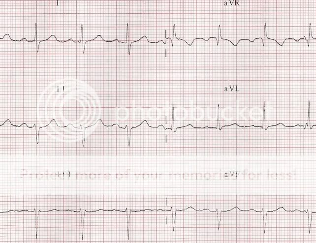
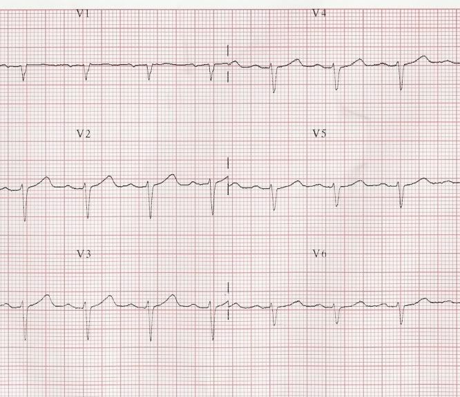
Well, what do we got here?
 

abuan

Forum Crew Member
43
0
6
i'm still learning ecg interpretation, but it looks like

normal axis sinus rhythm with a possible 1st degree av block and possible left bundle branch block with no st changes or ectopy at 90?
 
Last edited by a moderator:

emt_irl

Forum Captain
255
1
18
ive seen that rythm before in lead II i posted up about it also. from reading its like a bundle branch block of some degree or previous m.i
coming from a basic point of view

btw keep this thread up its a great idea
 

rhan101277

Forum Deputy Chief
1,224
2
36
ive seen that rythm before in lead II i posted up about it also. from reading its like a bundle branch block of some degree or previous m.i
coming from a basic point of view

btw keep this thread up its a great idea

I don't see any previous MI myself. I see pathological left axis deviation, I don't really see a BBB, but a hemiblock is present. Also there may be some electrolyte abnormalties.

Lets remember that everyone can interpret these different.
 

BLSBoy

makes good girls go bad
733
2
16
I see NSR with 1st Deg AV Blk.
 

DV_EMT

Forum Asst. Chief
832
1
0
To me, there's no sign of an MI. There's no ST elevation or depression noted in any of the leads. Looks like a Sinus Rhythm w/ 1st degree av block. The QRS is to short for it to be a Bundle Branch Block. I eyeballed it, looked like 0.10 seconds
 

TomB

Forum Captain
393
82
28
Lets remember that everyone can interpret these different.

That's true, but that doesn't mean they are all correct.

However, you are correct. This 12-lead ECG shows sinus rhythm with left anterior fascicular block.

Tom
 
OP
OP
Dutch-EMT

Dutch-EMT

BScN
97
1
8
10 points for TomB!

Conclusion:
P tops followed by a QRS complex, so it's a sinusrythm.
PQ-time, well what can we say about it?
Frequenty about 85bpm
Axis: I = mainly positive, aVF = positive, so it's a left axis deviation.

QRS: it seems wide, but is it BBB? No, let's look to the QRS in V1-V6.
The R-top progression seems slow in V1-V6. The S is deeper than normal in V6.
A lower intraventricular conduction (is it correct english?),the R wich doesn't show up very well, in combination with a left axis deviation and a deep S in V6...

Answer: Left anterior hemiblock or left anterior fascicular block!

Okay, next one!!!

34260674.jpg


For a bigger screen, click HERE
 

MrBrown

Forum Deputy Chief
3,957
23
38
Tooo easy mate! NSR (1500 method says 60bpm so technically is sinus brady) with anterior MI; I picked one of those up the other week.

How come Lead II in the first strip is negative instead of positive?
 

TomB

Forum Captain
393
82
28
And no chief complaint?

At first glance it's acute anterior STEMI vs. benign early repolariation.

I would capture another ECG in 5 minutes and compare (assuming signs and symptoms of ACS).
 
OP
OP
Dutch-EMT

Dutch-EMT

BScN
97
1
8
1mm ST-elevation mostly isn't significant... mostly...
I personally never make a MI out of this when it's the only sign i've got...
 
OP
OP
Dutch-EMT

Dutch-EMT

BScN
97
1
8
At this EKG the R is very small in V1 and V2, and a deep S. But that's normal.
I have a standard procedure to look at EKG's, in hope to catch all information out of it.

1. Frequency
2. Rhytm: is it regullar or irregular
3. P's: yes/no, are they looking the same? P's followed correctly by a QRS?
4. PQ time (<0,2sec), QRS time, BBB?
5. Heartaxis: Look at I and aVF
6. Hypertrophy? P in V1 (atrial hypertrophy) and S in V1 and R in V5 >3,5mV (LVH). R in V1 and S in V5 >1,05mV (RVH)
7. MI? Q's, ST and T
 

Scubamedic

Forum Probie
21
0
1
Frequency

1. Frequency
2. Rhytm: is it regullar or irregular
3. P's: yes/no, are they looking the same? P's followed correctly by a QRS?
4. PQ time (<0,2sec), QRS time, BBB?
5. Heartaxis: Look at I and aVF
6. Hypertrophy? P in V1 (atrial hypertrophy) and S in V1 and R in V5 >3,5mV (LVH). R in V1 and S in V5 >1,05mV (RVH)
7. MI? Q's, ST and T


I am a medic in the U.S and I understand your list except for the 1st word... frequency. 2-7 are simular to what I learned, but what do you mean by Frequency?

Here is a great site for anyone that wants to work on their 12 lead's, I study here among other websites.
http://library.med.utah.edu/kw/ecg/index.html
 
Last edited by a moderator:

8jimi8

CFRN
1,792
9
38
i think he means heart rate
 

Scubamedic

Forum Probie
21
0
1
Lol.

Thanks, that was my only guess. I got one to post I found. I had to get out my little als book and measure it, and I still got it wrong.
ecg_12lead-leftanteriorfascicularbl.gif
 
OP
OP
Dutch-EMT

Dutch-EMT

BScN
97
1
8
The EKG I posted was a totally normal EKG.
It's made on myself by the cardiologist during a cardiologyclass.
 
Top