rhan101277
Forum Deputy Chief
- 1,224
- 2
- 36
I had a call for a 83 y/o female with SOB. We get there she is not responsive but had adequate RR with diffuse rales and a hx of CHF and heart problems, previous strokes and MI's.
I put her on oxygen because I am getting sats of 65%. I then put her on the monitor and have 3rd degree HB at a rate of 20 and I can't hear a BP.
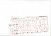
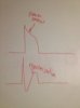
I pace at 70bpm/50ma and I feel I have got capture even though the QRS looks like "phantom QRS waves". Her b/p comes up and sats come up so treating the patient not the monitor. Still it appears that the underlying rhythm is marching through the paced QRS at where the absolute refractory period should be and this is impossible. My vitals signs increased and I checked the underlying rhythm several times during pacing and it was the same. Any thoughts? Normal paced rhythm has broader QRS and wide T waves, but I treated patient.
Thoughts?
I put her on oxygen because I am getting sats of 65%. I then put her on the monitor and have 3rd degree HB at a rate of 20 and I can't hear a BP.
I pace at 70bpm/50ma and I feel I have got capture even though the QRS looks like "phantom QRS waves". Her b/p comes up and sats come up so treating the patient not the monitor. Still it appears that the underlying rhythm is marching through the paced QRS at where the absolute refractory period should be and this is impossible. My vitals signs increased and I checked the underlying rhythm several times during pacing and it was the same. Any thoughts? Normal paced rhythm has broader QRS and wide T waves, but I treated patient.
Thoughts?