STEMI Mimic?

mrhunt

Forum Lieutenant
193
29
28
So 70 yo F, A&0X4 gcs15 cc of general weakness.

pt has extensive cardiac hx with multiple MI's and stents placed in prior years.
Pt has NO chest pain , shortness of breath, Nausea, vomiting or any other symptoms aside from weakness.
SP02 96% RA, Allergy to ASA, Systolic in 180's, Pulse 80's....

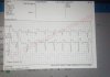
Flew out pt, MD consult concured this appeared to be an inferior stemi. Flight crew seemed to agree. Upon arrival to Stemi receiving facility flight crew took one last 12 lead and elevation was completely gone and STEMI alert cancelled.

did i **** up here?Was there something im missing that i didnt see?Im a new medic and still learning. Help me out here yo.

Basically just flew pt out, IV access, No nitro cause inferior involvement and Zero chest pain. No Aspirin cause of allergy. Could have done supportive 02 with mona protocol but SP02 fine and No pain to give fentanyl (we dont have morphine)
 

Attachments

  • 12Lead_29e0b63d-05ba-4b15-9984-7bf2effe0505__12Lead-0005.png
    12Lead_29e0b63d-05ba-4b15-9984-7bf2effe0505__12Lead-0005.png
    45.4 KB · Views: 265

DesertMedic66

Forum Troll
11,268
3,450
113
Elderly female patient with an extensive cardiac history with general weakness. Mix that with a 12-lead that has elevation in III and aVF with reciprocal changes. I would have done the same thing. This may have been a TSTEMI (Transient STEMI).
 
OP
OP
M

mrhunt

Forum Lieutenant
193
29
28
initial 4 lead showed obvious elevation in II and III.... `12 isnt QUITE as clear.

Are you seeing st elevation in lead 2 as well to complete the inferior?or is that just me?
 

Jn1232th

Forum Captain
359
81
28
I would of done same thing. Elevation is 3 and avf. You have reciprocal changes. History of MI. Sounds like a stemi alert to me. Did you do a V4R at all?
 

Jn1232th

Forum Captain
359
81
28
With being HTN I doubt any involvement. I would expect patient to be hypotensive instead. Other than getting a V4r I would have done everything the same
 
OP
OP
M

mrhunt

Forum Lieutenant
193
29
28
That's the initial 4 lead...
 

Attachments

  • 20191007_202415.jpg
    20191007_202415.jpg
    416.7 KB · Views: 209

StCEMT

Forum Deputy Chief
3,052
1,709
113
I'd have done the same thing based on this.

And go ahead and scratch MONA from the list of things you use.
 

DesertMedic66

Forum Troll
11,268
3,450
113
initial 4 lead showed obvious elevation in II and III.... `12 isnt QUITE as clear.

Are you seeing st elevation in lead 2 as well to complete the inferior?or is that just me?
I’m only on my phone and it’s not letting me zoom closely so I can’t tell if there is any elevation in lead II. If there is, it is marginal.

You technically only need elevation in 2 contiguous leads. So III and aVF is enough to call it.

Also keep in mind that the 4 lead is only useful for obtaining a rate and rhythm for EKGs. It has a lot of filters added so you can either have what appears to be a STEMI but once you record a 12-lead there will be none or vice versa.

Also as @StCEMT has said, MONA isn’t a huge thing that is advocated by anyone anymore. Fentanyl is preferred over morphine and oxygen should only be used to maintain an SpO2 >93%
 
OP
OP
M

mrhunt

Forum Lieutenant
193
29
28
cool! u learn everyday. thanks everyone. sounds like aside from v4R, i did the right thing. ......and even v4R isnt like a critical thing.... just would have been nice to remember....
 

StCEMT

Forum Deputy Chief
3,052
1,709
113
If you're in a pinch, you can compare STE. If III > II, that's a good sign of RV involvement. I work urban EMS, so the odds of me ever doing V4R when I have easy alternatives are basically 0. I believe it's actually a better method than V4R, I'd have to double check though.
 

Aprz

The New Beach Medic
3,029
664
113
To me, it is clearly not a STEMI, and I would've been comfortable not calling it one. There are several things about this ECG that tells me it is not a STEMI.

First, you need to consider the ratio between the size of the QRS complex and the ST elevation. Whenever you have a large R wave, you will have associated larger ST depression going the opposite way. The same is true for when you have a large S wave, you will have associated larger ST elevation. This is why it is considered normal and a STEMI-mimic in left ventricular hypertrophy "strain patterns." I like @TomB (I believe it was him) from ems12lead.com using a ratio of 0.2 (ie every 1 big box or 5 mm = up to 1 mm ST change OK). So example, in lead 3, we can see that the S wave goes off the 12-lead itself, but we can see that it is ATLEAST 4 big box or 20 mm away. That means up to 4 mm ST elevation would acceptable. We have 1 mm ST elevation... This is important to consider for BOTH small and large QRS complexes. So if you have very very small QRS complexes, but 0.5 mm ST elevation, that is significant and considered a STEMI! even if it doesn't meet AHA 1 mm in two contiguous lead criteria (that is a missed STEMI if you do not call it!).

The other thing you need to consider is that, in all likelihood, when the ST changes with T wave changes are facing opposite directions of the QRS, like the QRS and T/ST vectors are opposite directions, in all likelihood, you are dealing with a STEMI mimic. This is true with most STEMI mimics like paced rhythms, left bundle branch blocks, left ventricular hypertrophy, etc. So we can see her that the ST changes AND the T waves are facing opposite directions from the QRS. If you look at the machine's interpretation, the T wave is -47 degrees while the QRS is +78 degrees. I mean those are just numbers, but you can literally see it...If it has a deep S wave, then there is ST elevation with a positive T wave. If it has a tall R wave, then there is ST depression with a negative T wave. That is what I mean by the ST changes/T waves facing opposite of the QRS.

Although not a huge deal to me, but we see very small T waves as well, which is not normal in an acute event in my opinion. I mean in comparison to the QRS, these are baby T waves.

Anyways, if it was me, I wouldn't have activated STEMI alert, but I am overly confident and stupid, so that is just me. I would not knock anyone who activates this even if they were a veteran paramedic.

For the record, since somebody mentioned lead V4R, I am not really a fan of it. You can tell enough with comparing lead III and II ST changes and looking at ST elevation in lead V1 and/or ST depression in lead V2 (reciprocal change) to tell if there is significant right ventricular involvement, and really, the inferior wall MI being sensitive to Nitroglycerin is kinda bogus (https://www.ncbi.nlm.nih.gov/pubmed/26024432). It is more important to look at your margin of error with blood pressure, how much it will drop/tank if you give it to them rather than what type of infarction it is. An anterior or lateral wall MI with no signs of significant ventricular involvement can tank as well. That being said, I will give Nitroglycerin if the blood pressure is high, but if it is close to normal to low, I will withhold it.

I tagged Mr Tom B so he is free to call me out. I can also tag @Christopher (also from ems12lead, created that Google survey/test thing that was popular for comparing STEMI vs STEMI mimics).

Edit: @StCEMT You beat me to saying lead III vs II, but I'll raise you lead V1 and V2!
 

Gurby

Forum Asst. Chief
818
597
93
To me, it is clearly not a STEMI, and I would've been comfortable not calling it one. There are several things about this ECG that tells me it is not a STEMI.

First, you need to consider the ratio between the size of the QRS complex and the ST elevation. Whenever you have a large R wave, you will have associated larger ST depression going the opposite way. The same is true for when you have a large S wave, you will have associated larger ST elevation. This is why it is considered normal and a STEMI-mimic in left ventricular hypertrophy "strain patterns." I like @TomB (I believe it was him) from ems12lead.com using a ratio of 0.2 (ie every 1 big box or 5 mm = up to 1 mm ST change OK). So example, in lead 3, we can see that the S wave goes off the 12-lead itself, but we can see that it is ATLEAST 4 big box or 20 mm away. That means up to 4 mm ST elevation would acceptable. We have 1 mm ST elevation... This is important to consider for BOTH small and large QRS complexes. So if you have very very small QRS complexes, but 0.5 mm ST elevation, that is significant and considered a STEMI! even if it doesn't meet AHA 1 mm in two contiguous lead criteria (that is a missed STEMI if you do not call it!).

The other thing you need to consider is that, in all likelihood, when the ST changes with T wave changes are facing opposite directions of the QRS, like the QRS and T/ST vectors are opposite directions, in all likelihood, you are dealing with a STEMI mimic. This is true with most STEMI mimics like paced rhythms, left bundle branch blocks, left ventricular hypertrophy, etc. So we can see her that the ST changes AND the T waves are facing opposite directions from the QRS. If you look at the machine's interpretation, the T wave is -47 degrees while the QRS is +78 degrees. I mean those are just numbers, but you can literally see it...If it has a deep S wave, then there is ST elevation with a positive T wave. If it has a tall R wave, then there is ST depression with a negative T wave. That is what I mean by the ST changes/T waves facing opposite of the QRS.

Although not a huge deal to me, but we see very small T waves as well, which is not normal in an acute event in my opinion. I mean in comparison to the QRS, these are baby T waves.

Anyways, if it was me, I wouldn't have activated STEMI alert, but I am overly confident and stupid, so that is just me. I would not knock anyone who activates this even if they were a veteran paramedic.

For the record, since somebody mentioned lead V4R, I am not really a fan of it. You can tell enough with comparing lead III and II ST changes and looking at ST elevation in lead V1 and/or ST depression in lead V2 (reciprocal change) to tell if there is significant right ventricular involvement, and really, the inferior wall MI being sensitive to Nitroglycerin is kinda bogus (https://www.ncbi.nlm.nih.gov/pubmed/26024432). It is more important to look at your margin of error with blood pressure, how much it will drop/tank if you give it to them rather than what type of infarction it is. An anterior or lateral wall MI with no signs of significant ventricular involvement can tank as well. That being said, I will give Nitroglycerin if the blood pressure is high, but if it is close to normal to low, I will withhold it.

I tagged Mr Tom B so he is free to call me out. I can also tag @Christopher (also from ems12lead, created that Google survey/test thing that was popular for comparing STEMI vs STEMI mimics).

Edit: @StCEMT You beat me to saying lead III vs II, but I'll raise you lead V1 and V2!

:eek:
 

Tigger

Dodges Pucks
Community Leader
7,844
2,794
113
If you have a changing EKG, something is happening.

I don't know if there are reciprocal changes in I and AVF. Are they reciprocal changes or the expected morphology from a right bundle (the QRS is wide but it is difficult to see what the morphology in V1 is)? The ST segment in the inferior leads is concave which is less suggestive of STEMI. The patient also does not have chest pain. I really do not want to make a call on this patient. If through serial 12 leads I noted changes, I probably would.

It would be interesting to see what her troponin was and whether she received a less emergent cath.
 
OP
OP
M

mrhunt

Forum Lieutenant
193
29
28
I have to mention we took three 12 leads all showing Stemi Inferior but i know you mean serial 12 leads over a longer period of time and not back to back....
 

Tigger

Dodges Pucks
Community Leader
7,844
2,794
113
I have to mention we took three 12 leads all showing Stemi Inferior but i know you mean serial 12 leads over a longer period of time and not back to back....
Honestly the above EKG could be the baseline EKG. The point being is that you know it is not baseline if you have obvious changes.

Also as mentioned, 4 lead traces cannot be used to evaluate for ST changes. You don't have to implement care protocols if they aren't appropriate for the patient. Giving oxygen and morphine here is not appropriate.
 
OP
OP
M

mrhunt

Forum Lieutenant
193
29
28
I dont entirely understand your statement.......I dont know what her "normal" ekg looks like.....Just what i took on scene and followed protocol and my best judgement as a new medic.

I didnt see any evolvement of st elevation because the 12 leads were taken minutes apart or less....
 

Aprz

The New Beach Medic
3,029
664
113
Like I said above, it's not a STEMI, I described my reasons why above (https://www.emtlife.com/threads/stemi-mimic.48053/#post-681280). That's OK. Nobody is going to knock you for doing what you think was right and trying to follow your protocols.

There is the patient's baseline and then your baseline that you took. If you do serial 12-leads, you can see if the next 12-lead deviates from the baseline set that YOU took.

When you put the patient on the monitor, either a 3-lead or 4-lead, it is usually called "monitor mode". When you do a 12-lead, it is usually called "diagnostic mode". They use different filters that can hide, create, or reveal ST changes. It is possible to put the patient on the 3 or 4 lead, see ST changes, but for those ST changes to go away when you do a 12-lead. At the same time, it is possibly to put the patient on the 3 or 4 lead, not see ST changes, but for ST changes to be revealed when you do a 12-lead. Usually though, if there is ST elevation or lack of, it'll be the same for the 12-lead. It's possible for there to be a difference in diagnostic and monitor mode, something important to note, but it is not usually the case. Christopher Watford wrote all about it here http://ems12lead.com/2014/03/10/understanding-ecg-filtering/, but most of it goes above my head.
 

Summit

Critical Crazy
2,691
1,312
113
Patients can have transient ST elevation. Pt might still get the stat cath, especially with an EKG like that, if they have enzymes. But with the ST elevation gone, cancelling the alert gives time to adequately work up an unclear situation so that the receiving facility isn't pressured by door to balloon time for a STEMI.

In ED/ICU we'd do continuous 12 lead monitoring on a patient like this on our MX800/X2 either with the full 12 lead set or more often with the EASI 12 lead interpolation placement and it is an indication to actually turn on the STe alarms (normal disable to avoid alarm fatigue) to catch transient elevation.

Did you ask what the trops were?

Did they do a right sided ekg and 15 lead?
 
Last edited:
OP
OP
M

mrhunt

Forum Lieutenant
193
29
28
We typically dont get follow up information on pt's. Especially when taken to main STEMI Centers outside of our area unfortunately.
 
Top