EKG Strip: Progression of ST Elevation

jedirye

Forum Lieutenant
114
0
0
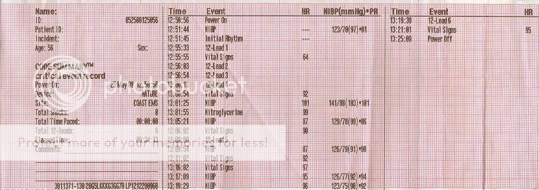
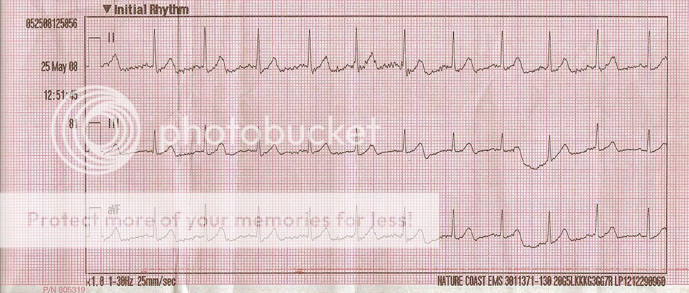
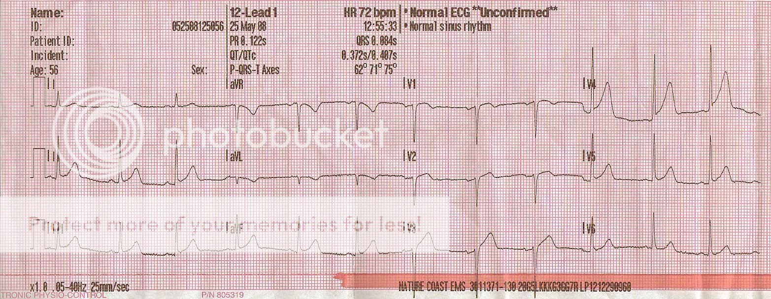
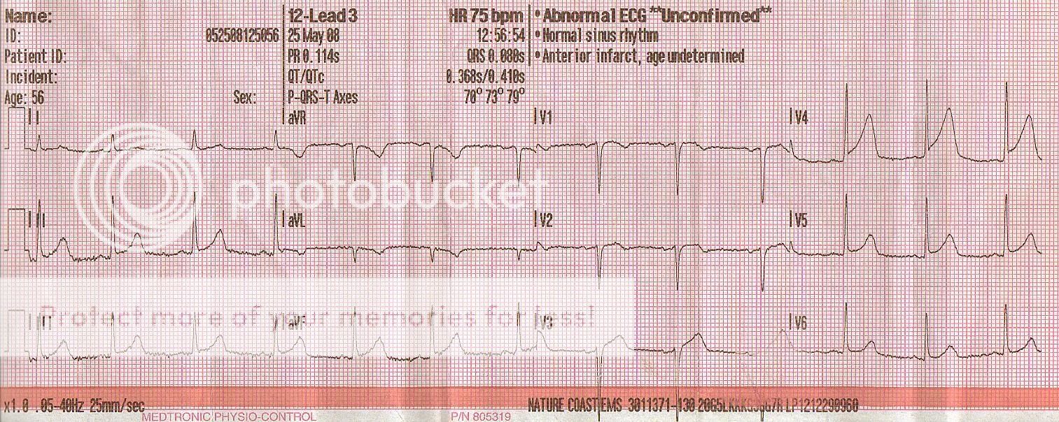
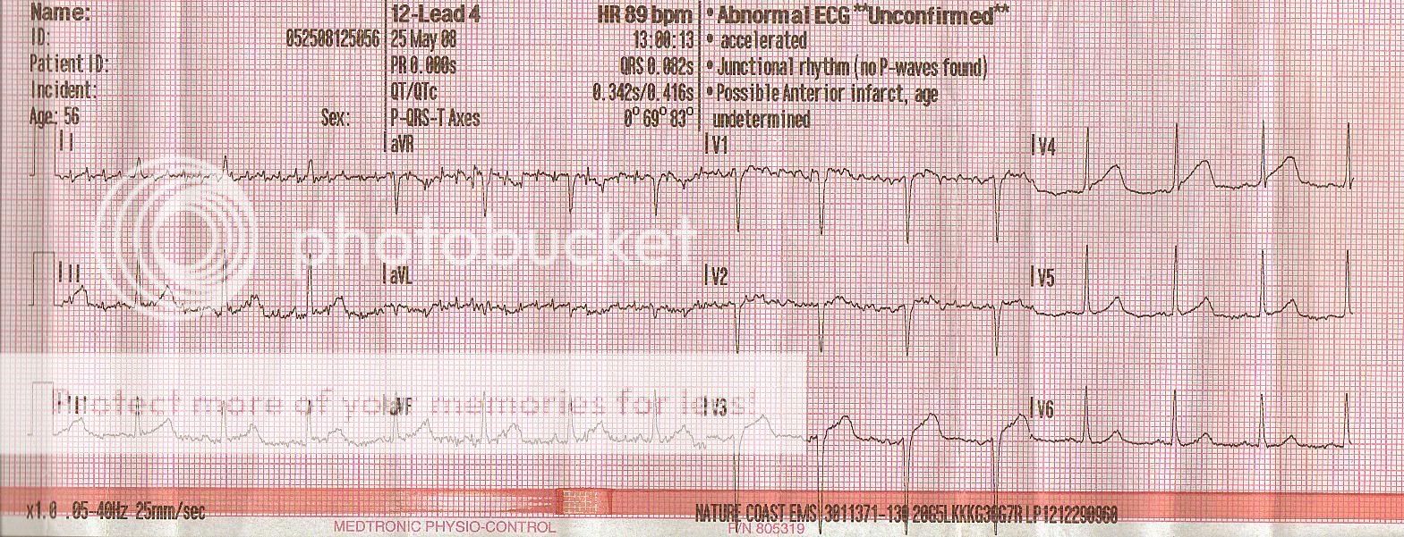
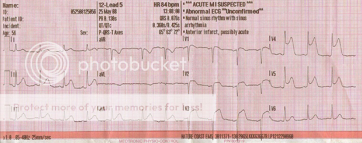
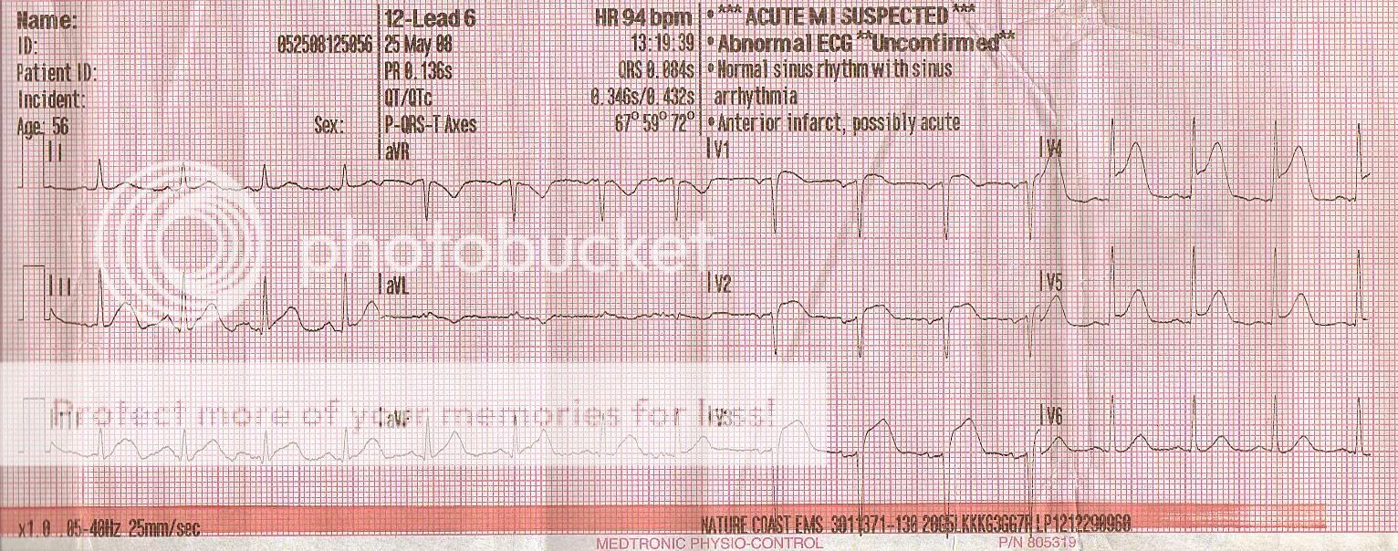
I found this strip while re-reading my EKG book, I guess I was using it as a bookmark or something. I have about 50 other strips scattered about in various locations, I'd like to scan them all but it is terribly time consuming. Figured I would share a cool strip as you can see the progression of ST elevation (hence title). As you can see from the date, it was about a year or so ago so I am not familiar with the story or treatment so take it for what you may. I found it quite interesting to note the time line of the 12-leads and the importance of repeat 12-leads...

Scan1.jpg


Scan2-1.jpg


Scan3-1.jpg


Scan4-1.jpg


Scan5.jpg


Scan6.jpg


Scan7.jpg


Scan8.jpg


-rye
 

MSDeltaFlt

RRT/NRP
1,422
35
48
Drastic change in 8 minutes. Impressive.
 

artman17847

Forum Crew Member
83
0
0
great set of 12 leads! i'am really curious as to how this pt presented and what TX's were given. you can clearly see the ST elevations in the anterior chest leads, would have been a good case to move a lead to the back to get a posterior look. thanks for posting.
 

Ridryder911

EMS Guru
5,923
40
48
Ironically, I did not see any reciprocal changes.

R/r 911
 

maxwell

Forum Crew Member
68
0
0
Classic anterior wall MI (they come on right quick!) Lest we forget the septal changes (they're not as dramatic, though) - but they're still mighty concave. Excellent set of tracings. Hope the pt. did well.
 

VinBin

Forum Captain
274
5
18
Just a quick question for the older medics, in your experience how prominent do Q-waves (with no other EKG abnormalities) have to be for you to consider them significant or at least keep on you on "alert"?

I have always heard the Q-wave being 1/3 of the amplitude of the QRS complex being the standard for indicating injury to the muscle I have also seen what would be considered physiologically normal Q-waves in acute MI's with little to no associated ST changes.
 

maxwell

Forum Crew Member
68
0
0
Just a quick question for the older medics, in your experience how prominent do Q-waves (with no other EKG abnormalities) have to be for you to consider them significant or at least keep on you on "alert"?

I have always heard the Q-wave being 1/3 of the amplitude of the QRS complex being the standard for indicating injury to the muscle I have also seen what would be considered physiologically normal Q-waves in acute MI's with little to no associated ST changes.

Q waves don't get me (or most cardiologists, even) pressed. STEMIs do. 1/3 of the amplitude is a good rule to go by. Often times after blood flow spontaneously restores itself ST segments normalize and they can (usually do) get Q waves in the corresponding leads - often times we call this being "Q'd out" (when it's too late to cath 'em).
 
OP
OP
jedirye

jedirye

Forum Lieutenant
114
0
0
Just a quick question for the older medics, in your experience how prominent do Q-waves (with no other EKG abnormalities) have to be for you to consider them significant or at least keep on you on "alert"?

I have always heard the Q-wave being 1/3 of the amplitude of the QRS complex being the standard for indicating injury to the muscle I have also seen what would be considered physiologically normal Q-waves in acute MI's with little to no associated ST changes.


Ironically I have another strip with significant Q waves (as in, greater than 1/3 amplitude of QRS complex) and I think there was also T wave inversion. I have been meaning to post it as it was last shift and I was wondering the same thing concerning Q waves: If no elevation but Q waves and active chest pain, go code 2? No elevation, Q waves, and t wave inversion, code 2? I realize it's not just the 12-lead but the patient, patient's response to tx (asa, nitro, morphine, etc) but all relative to each other. Ah well, this will be another thread another time, perhaps...

-rye
 

Melbourne MICA

Forum Captain
392
13
18
Onward and Upward

Nice demonstration of the progression of the ECG changes in infarct. Also demonstrates the old addage "infarcting" not "infarcted". There looks to be inferior involvement as well so R and L coronary structures. Multi vessel disease but not a global infarct. V5 is great to watch as you scroll down the page and see the T wave rise, then the segment rise after it.

The reverse is interesting when you see resolving coronary artery spasm.

Thanks for the great ECG

MM
 
Top