What do you think this is??? (12 lead)

Grady_emt

Forum Captain
Messages
301
Reaction score
0
Points
0
Just looking to see what you guys think this rhythm (esp the initial one) is. I havent a clue, and we got totally different answers from each doc we asked.:unsure:

So on the last night of a long weekend, approx 0345 hrs my partner and I were dispatched to a Code 1 "Diff Breathing, Severe Resp. Distress/Cardiac Hx" with a BLS engine (we are ALS). Upon arrival, 54 yof pt found lying on left side in bed, and looks like crap (note, we have run this pt several times, normally very talkative and alert) Hx: Acute Renal Failure/Dialysis Pt, Diabetic, HTN, ^cholesterol, MI in 2001. Per roomate, pt is M/W/F Dialysis pt and last went on F of last week, and has been non-compliant with any of her meds x 3 weeks, which are the usual dialysis pt full-grocery-bag variety.

She looks like crap, resp approx 34-26, pulse 46 and thready, pressure 136/80, pulse ox 84 on room air. While awaiting fire for extrication assist from 2nd flr, pt placed on NRB, and the monitor was applied to show this...
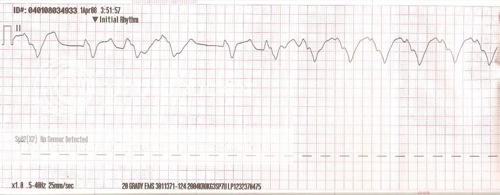
hypercalemiaPBriggs4108.jpg


Pt is non verbal except "need some oxygen" over about 6 breaths. extricated to unit, 12 lead applied to show
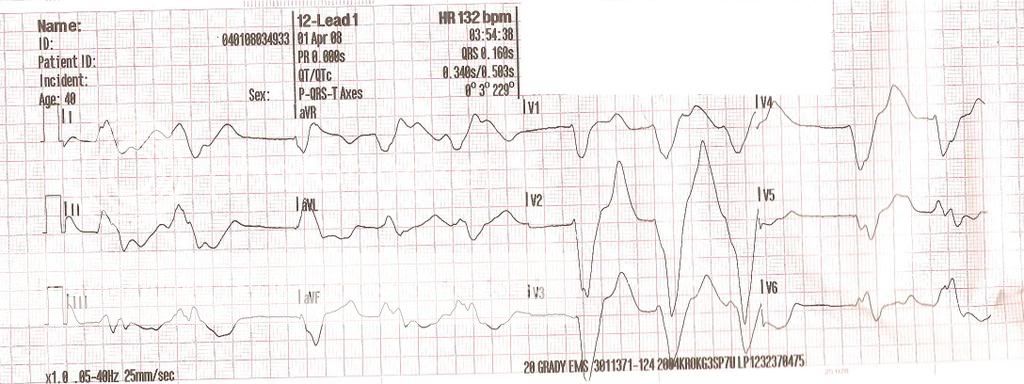
hypercalemia12lead1PBriggs41intrepr.jpg


Breath sounds are clear, - edema noted, pale mucosa, poor skin turgor, unable to establish IV (everythings all scarred over, EJ is completely flat). One more 12lead before leaving scene shows
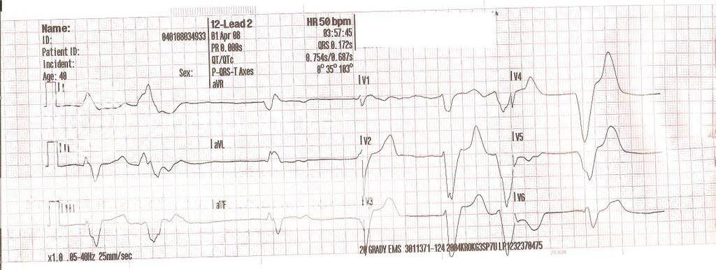
hypercalemia12lead2PBriggs41intrepr.jpg


We had about a 5 minute transport to ER, where pt went into v-fib that was defib-ed into Torsades, that after 4g MagSulf, 2xBiCarb, 2xEpi, 2xAtro, Calcium Gluconate, and Calcium Chloride, and D50, pt was again defib-ed into a sinus-ish rhythm with a pulse that she maintained for about 10 minutes before degrading back to v-fib which became her terminal rhythm.:wacko:
 
First strip accelerated IVR, commonly seen in electrolyte imbalances. Basically, they do not know their dead yet...

R/r 911
 
the last earthquake that hit LA?
 
:censored::censored::censored::censored:e. Gotta love a pt like that...the least they could do is make it a bit easier on you. Which they sort of did...:D

Rhythm aside, what was actually wrong with this person, and (if you'd been able to get a line) what would your treatement (if any) have been? (I'm guessing that you know what the problem was...pretty clear at this point...still a good learning point for some people though)
 
All are accelerated IVR. The rhythm and the first ECG also have multiple Premature IVR beats as well, whereas the last one has only 2 Premature IVR beats and is slowing down. It was slowing down for a reason.

Rid's right. Usually the cause of an electrolyte imbalance.

I believe the root cause of this pt's demise is the noncompliance with her meds causing an electrolyte imbalance which deteriated to an irretractably terminal rhythm.

And for an opinionated side note: Doctors give you prescriptions for a reason. They tell to change your lifestyle for a reason. They tell you to stop doing "this" and start doing "that" FOR A REASON.

Opinions are like buttholes. Everybody's got one. Most of them stink, and this one is mine. Just thought I'd throw it out there.
 
gonna have to agree with the others, first was an accelerated idioventricular, the fact that you said dialysis pt. shoulda summed it up, i hate ECGs from them. Their electrolytes are all out of whack, making their heart do some crazy stuff. She's obviously got some hyperkalemia issues or something, not at all surprised she coded on you.
 
First strip accelerated IVR, commonly seen in electrolyte imbalances. Basically, they do not know their dead yet...

R/r 911


This is what the attending at our hospital said "Thats the rhythm that you go into just before you die...that patient's not here are they?:o??"

He also said that it would most likely secondary to Hyperkalemia, and without seeing the pt, only the EKG, it was most likely irreversable (as it was).
 
As a preceptor told me, "Lead II-you have no clue"..:(..well that's as far as I've been into ECG so far....

......but I do recognize the accelerated IVR.

Thanks for posting up the strips....

-Matt
 
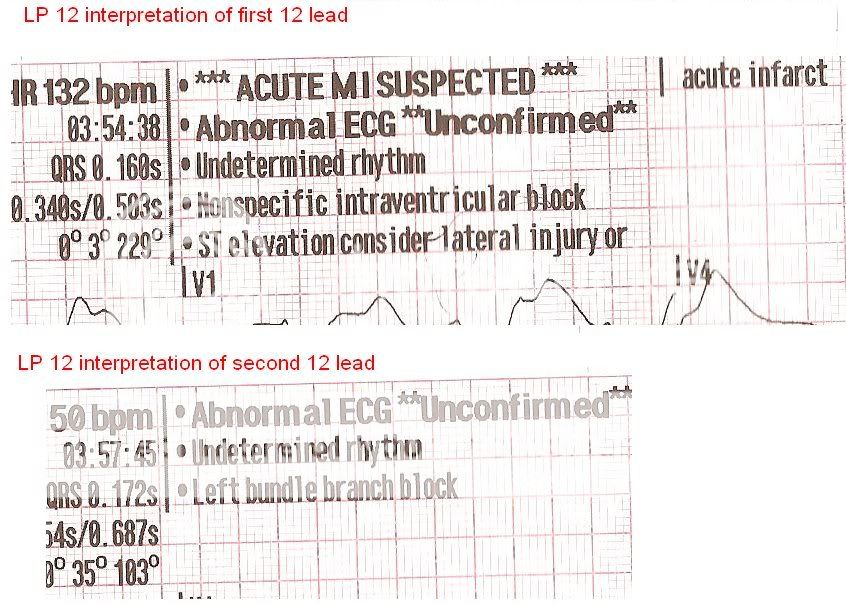
Just for kicks and giggles, here is what the LP12 showed for its interpretation of the 12leads, I had cut these out of the original posts. Notice that it says "Undetermined Rhythm", I thought that was kind of amusing and shows that you cant trust the machine, you need to know how to read the 12lead yourself too. But above all, treat the patient!!!

interpretationof12lead.jpg
 
Ha, that'd be a reroute to a cath lab in LA and Orange County (machine interpet only. There's a classic published quote if someone wants me to post it).
 
Lets just say that it is one of those "strips" you dont want to see in the exam!!

I am glad that you mentioned the history and presentation of the patient, so that one can treat the strip and not only diagnose it. A lot of the i get a strip shuved in my hand and asked to treat it. My first question is then: "Was the patient symptomatic?". if the answer is: no, then my teatment is supportive and transport asap.

This patient was ill, and your treatment was good. When you guys IVR, is it: Idio Ventricular Rhythm?? I would go with accelrated idio ventrular for this patient. As for the Left Bundle Branch Block, i don't agree with the analysis of the 12 lead. Left & Right BBB is diagnosed in V1 & V6, not ( or only) in V2, but i can see why the machine did it.

Sorry about the loss of the patient.

I think the moniter was just spitting out phrases on this patient. Once we determined that we were not getting any type of IV access on this pt, our treatment consited of EKG, O2, and a Diesel bolus down the road to the ER since we were less than 5 out, there is no sense staying and trying things onscene (and it was a good thing, less than 5 minutes in the ER, she arrested).

This was actually our 4th arrest in two days. This was one, another was a respiratory rate of 6 we tubed that progressed to a full arrest while transporting(79yof cx, pronounced in ER after a bit) from a nursing home. Staff quote during our questioning "I have other people to go see, can't you just get her out of here?"

Another non-complaint dialysis pt severe pulmonary edema, when tubed the fluid came shooting out, evidently arrested in the ER after we left (46yof, non complaint, 4 blocks from ER, given 80lasix, NTG paste while awaiting fire for extrication from 4th floor.

Yet another Dialysis pt that I will post strips and scenario on later (+ maintained ROSC)
 
Sounds like a good call for IO.

I like the FAST PYNG especially when the pt is flat. I would like to try the EZ IO, however understand obesity can cause problems, although who ever see's an obese pt.

I was taught not to read the print out. This can bite you however if you miss the "suspect leads reversed" warning.
 
Is anyone thinking hyperkalemia? Pointy T waves, widened QRS, history of acute renal failure?
 
hyperkalemia...no, hypo is a maybe though. i don't belive that it is IVR since there are no U wave(which don't always have to be presents) and the fact that there is atrial respone in the P-waves. Torsades de Pointes makes more sense since the top of the QRS complexes twist the amplitude changes on the R wave.
 
hyperkalemia...no, hypo is a maybe though. i don't belive that it is IVR since there are no U wave(which don't always have to be presents) and the fact that there is atrial respone in the P-waves. Torsades de Pointes makes more sense since the top of the QRS complexes twist the amplitude changes on the R wave.


IVR does not always have U waves as well as there are no defined P waves either rather than a typical dying heart pattern with occasional an occasional atrial P wave (post 3'rd degree). Torsades de Pointes, would not be considered for several reasons. The QRS is too wide as well as the ventricular rate is too slow for ventricular tachycardia (remember Torsades is V-tach arising for multiple foci in "twisting or rotating" pattern). Although, the etiology of Torsades is usually caused from electrolyte imbalance.

R/r 911
 
.... from a nursing home. Staff quote during our questioning "I have other people to go see, can't you just get her out of here?"

A little off topic.. but, typical nursing home
 
My BLS interpretation? This Pt. is screwed

O2 AED at the ready ( unless valid DNR ), monitor vitals and run like hell for the ER
 
I would go with an IVR with a sine wave pattern. I don't see any P waves and the QRS complex is wide - the likely cause being increased serum K+ levels. The patients medical history, more specifically the recent history confirms the rest. I would be in a hurry to establish vascular access and start administering Calcium Chloride and Sodium Bicarbonate - it's the only way your going to turn this around (if it can be done - not likely in this case).
 
Back
Top