Name the Rhythm: 52 year old male

As for treatment....... Well, I know you didn't ask for our treatment. But obviously one would need a lot more to know what our treatment would be.


My interpretation is just based off the ECG print out and the age/CC

My bet would be fluid challenge and then beta blockers if necessary.
 
Slight hints of a WPW. Not saying it is, but could see the argument, look at lead 3.

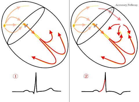
For WPW consider that the delta wave is formed by fusion of wavefronts from two separate impulses. (1) impulses pre-exciting a part of the ventricles which then meet (2) impulses using the normal conduction system.


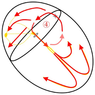
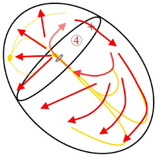
In all (regular) narrow complex tachycardias involving an accessory pathway there is no "pre-excitation" occurring as the AP is only used in a retrograde manner.


Even still, when the AP is used in an antegrade fashion, there is also no "pre-excitation" as the AVN is the retrograde pathway and there is no fusion of two wavefronts. However, in this case a wide complex will be present as the complexes are formed from slow traversal thru the ventricular myocardium until it reaches the conduction network.


So unless you've got an irregularly irregular rhythm like AF w/ WPW or Atrial Flutter and WPW, you will not see Delta Waves during the tachycardia because there is no fusion of two wavefronts.
 
Treat the patient not the monitor?

I'm going to beat you with a LP5 or worse assign you to the truck that never gets off shift on time, this is rhythm interpretation not an alphabet soup class ;-)

The answer to this case lies in following the steps of rhythm interpretation, taking into account information in all 12 leads.
 
I'm going to beat you with a LP5 or worse assign you to the truck that never gets off shift on time, this is rhythm interpretation not an alphabet soup class ;-)

The answer to this case lies in following the steps of rhythm interpretation, taking into account information in all 12 leads.

Haha sorry had to say it
 
I'm still going with a flutter. Thought I saw some "blips" of p waves some buried in the t waves
 
Cardiovert for svt (too soon?).

Looks like a narrow-complex regular tachycardia with normal sinus activity. All the other schmutz (ST segments, T wave morphology, P wave appearance) seem like rate-related or artifact, and just don't look so interesting.

Without any other clinical data, I'd put a liter of NS on a pressure bag and keep on talking with him. See what comes up.

Then shock him again (can't resist!).
 
Yes and as long as pt is stable I would push adenosine, then maybe I could make out p waves a little better.
 
Ah, you were catching on that I was posting name that ECG here.

Rate? 167-187. I trust the monitors HR which is 176.

Regularity? Regularly regular

P-waves? Yes. I think it's best seen in lead I. I can see it in V4-V6. Would be questionable in V2-V3, but with the other leads, I would say those notches at the end of the T-waves aren't artifacts, but p-waves superimposing on the T-wave making a notch.

256u2dz.jpg

Arrows pointing at notch.

PR interval? Hm, not sure if it's consider calculable. I decided to use lead II. The second half of what I would call the T-wave is very tall (like when there is LAE or RAE, since both atrias are depolarizing at the same time and one has more surface than the other, it makes a tall p-wave; I know people say p-pulmonale, the tall p-wave >2.5 mm is RAE, but I believe I read that it's non specific for which one it is per Chou's book in the first few chapters I read (haven't read the whole thing). I am applying the same idea, but with a T-wave instead). I would say in II, it looks like 0.12s. Looking at the other leads, it's like 0.08s, but like what Garcia's book said, it's the longest that counts.

123v47p.jpg

Arrow pointing at notch, all of the sudden it gets bigger cause of combined electrical activity.

QRS duration? 0.04s

Rhythm DDx? Well, it's obviously an SVT, and I just happened to read from ems12lead the different types of rhythms and SVT can be: sinus tachycardia, atrial fibrillation, atrial flutter, atrioventricular nodal re-entry tachycarda, accessory pathway mediated tachycardia, atrial tachycardia, multifocal atrial tachycardia, and junction tachycardia. Sinus tachycardia is a possibility. It's regularly regular so not atrial fibrillation. I would expect to see flutter waves between the T-wave in the inferior leads since there is some gaps in there (and atrial flutter is usually best seen in the inferior leads and pointy T-wave in V1 I recall). AVNRT is still new to me so I am not sure. Also with accessory mediated, I think it's talking about WPW, and I don't see any delta wave (I try not to look too hard at the 12-lead, lol) and if I am correct about the PRi then no. Atrial tachycardia is possible if I am incorrect about the PRi and whether the P-waves are positive/negative. Where I am seeing P-waves, it looks positive in I, II, III, and aVF (makes the T-wave peaked). It's regularly regular so not multifocal atrial tachycardia either. It has a P-wave so it's not junctional tachycardia either. I think it's sinus tachycardia even though it's questionable with that rate.

35jcw1j.jpg

This is in regard to me not seeing any flutter waves in the inferior lead.

Axis? QRS: I is positive, aVF is positive. This is in the normal axis. If you use the I, II, III trick, I is positive (it's going to the patient's left), II and III are positive (it's going down) so it's going down and to the left, which is normal. aVL is most isoelectric/equiphasic so I would say around +60 degrees. Z: V3-V4 are equiphasic so +20 - +40. Normal R-wave progression.

QTi/QTc? Once again, I am not sure if considered calculable. Since the atria is depolarizing almost simultaneously as the the ventricle is repolarizing, I'd say to assume that calculating it even with the P-wave inside of it is OK. QT is about 280 ms. QTc is about 479 ms (280/((60/176)^(1/2), I trusted the monitors HR).

ST/T-wave changes? I don't see anything in the inferior leads (that have a gap before the T-wave) It's difficult to tell since I really don't a good TP segment to use and going off of the PRi instead. In tachycardias, sometimes it looks like there is ST depression because the atrial is repolarizing after the ventricle depolarize I think. I don't think the P-wave in V1 could be negative enough to make it look like retrograde T-waves so maybe retrograde T-wave in V1, but that's all.

Overall DDx? No (bad word)ing clue. :D Dehydration caused by something like an infection is all I'm thinking. If that's the case, fluid resuscitation would be better than administering adenosine since this tachycardia is compensating/terminating the rate without more fluids would be harmful. I cannot think of anything else that I would associate with this 12-lead.
 
Last edited by a moderator:
Why give your own diagnosis when Aprz does it for you?

It's like you read my mind Aprz :)

This is sinus tachycardia at ~180, P-waves are best visible in the anterior precordials (V4-V6). In those leads a PRi of ~160ms is seen, which transfers well to the limb leads. Once you do so you'll notice really tall peaky P-waves which look like T-waves.

I'd run this case on EMS 12-Lead back in May, so I changed the story here to make it hard to search for since all I wanted was a rhythm interpretation :)

If you were wondering how the crew treated it, well they presumed it to be a narrow complex tachycardia of uncertain etiology. They gave a fluid bolus and then adenosine and found all that happened was it ramped right back up to nearly the same speeds as before, albeit this time with easier to see P-waves. Stimulant usage was thought to be a factor.

I like to practice rhythm interpretation as a cold read, ignoring treatments and focusing on differentials. It helps you keep your eye on the prize, which is arriving at the short list of probable differentials. You can then weight them against your clinical findings and formulate a treatment plan.

Sticking to the rules as Aprz did is a wonderful way to ensure you don't miss anything. This case has upright P-waves in the limb leads with a normal PRi, which excludes reentrant causes.

Do I think this case is one where adenosine is reasonable? As long as it is logically arrived at and crews don't start rebolusing it, or moving to calcium channel blockers, or cardioversion when it doesn't work.

On a more advanced note, what's almost as important as a tachycardia is how it starts and how it stops! Capturing behavior during and after adenosine can hold a wealth of information.

Ramping up and down is a feature of automatic tachycardias, which are not likely to be solved by adenosine and, depending on the cause, may not to be solved with cardioversion.

I hope you guys and gals enjoyed this case as much as I did when I first saw it!
 
Last edited by a moderator:
Thanks! Been a big fan of a lot of the blogs including yours and ems12lead and been practicing a lot of 12-leads on ECG Wave Maven like you mentioned, EKG Club on Facebook, and just solved the 100th 12-lead in ECGs Emergency Physician Part 1 yesterday. Glad to know that my work has been paying off. :)
 
Back
Top