Electrophysiology behind Commotio Cordis?

OnceAnEMT

Forum Asst. Chief
Messages
734
Reaction score
170
Points
43
I have always been under the impression that commotio cordis was caused by an impact to the chest wall that was juuuust right in order to essentially "create" an R-on-T, resulting in vfib. For whatever reason the electrophysiology of the event was brought up in an AT class the other day, and the instructing GA didn't seem 100% on what he was saying about it, so I did some research (I don't remember what he said now). I'm reading that the impact occurs during the ~30milliseconds of the uphill of the T wave. This makes sense, as it would interrupt the repolarization of the ventricles, thus introducing vfib. But, what does this look like on an EKG?

I suppose my overall question is, could someone briefly explain the "why" behind commotio cordis? And if possible, describe what an EKG would show? (Yes, yes, I know we wouldn't see anything but a (hopefully) shockable rhythm on this kind of patient, but lets assume that patient happened to be wearing a Holter monitor or was hooked up to your LP when a baseball so perfectly entered through your back window).

Thanks guys.
 
Mechanical force can impart an electrical charge! Percussive pacing is actually a thing, where you strike the chest repeatedly to pace a patient. (don't actually do this)

Mechanical stretch causes changes in many ion channels (K+ and Ca2+ being those with the largest influence), which in turn generates an electrical potential. They're not 100% certain which contribute the most in commotio cordis, but they've found if they block potassium channels that mechanical pressures affecting stretching of the myocardium prevents VF formation. This was in studies of pigs, so take that in stride.

That time period about 40 ms prior to Tpeak is a period when repolarization dispersion is maximal. What does that mean?

Let's start with depolarization. Your heart depolarizes in a very orderly, triggered process. It is a traveling wave.

Repolarization is not an orderly process. Repolarization isn't triggered. Repolarization is not a wave. It has no real "front". Each individual myocardial cell repolarizes on its own time. In fact, repolarization occurs in the opposite order from depolarization. Depolarization starts in the endocardium while the first cells to repolarize are in the epicardium. If you were to look at the QRS complex of an individual myocardial cell it would have inverted T-waves. This begs the question: why are T-waves upright on a normal ECG? Because repolarization starts in the well fed epicardium rather than where depolarization began. To make things more interesting, the mid-myocardial cells (M-Cells) which make up the chewy nougat center of the myocardium, are the last to repolarize.

Back to the question, what does repolarization dispersion mean? Repolarization dispersion is the difference in timing between when the areas of the heart repolarize and are ready to accept a new impulse. If you have a wide repolarization dispersion, you have areas of tissue which are ready for a new impulse. A properly timed extrasystole (either a PVC generated intrinsically or a PVC generated extrinsically) can exploit this "heterogeneity". It starts new depolarization in the areas ready which end up creating tiny waves around the tissue which is not ready, only to end up reentering the later repolarizing tissues. This reentry can either continue as VT, PMVT, or degenerate into VF.

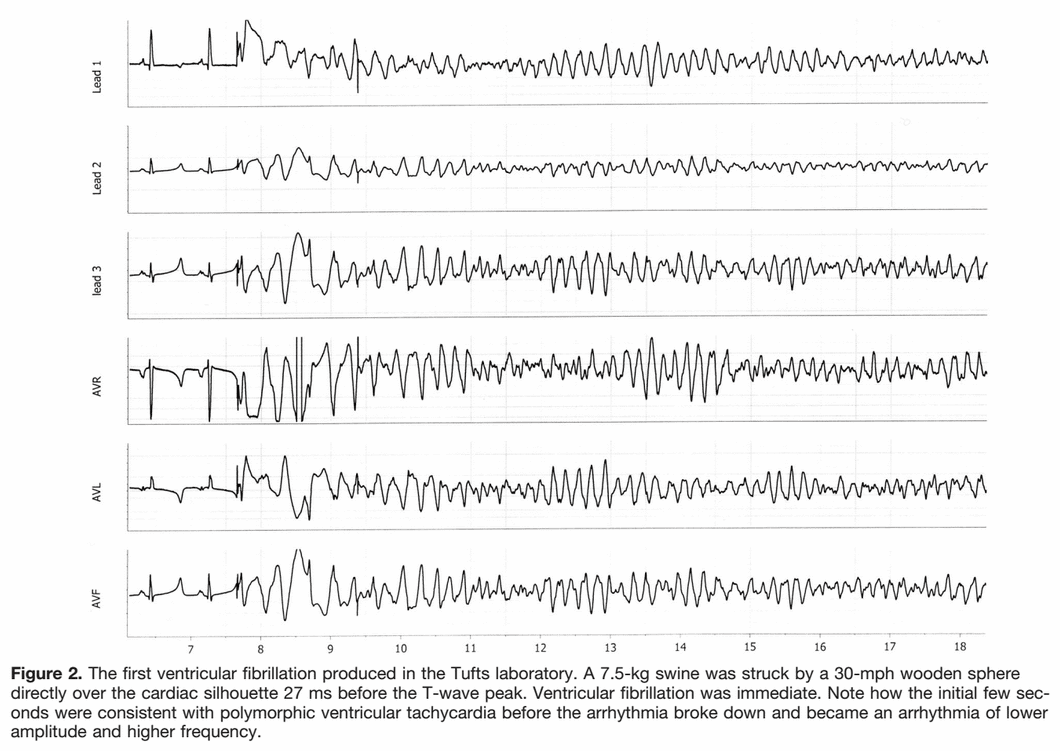
So back to commotio cordis. Mechanical impacts which generate spontaneous depolarization look nearly always like PVCs, often with LBBB morphology due to the section of the heart closest to the chest wall. So what would commotio cordis look like? Basically like R-on-T PVCs starting VF.
vf-commotio-cordis-impact-site.png

vf-commotio-cordis-initiation.png
 
Chris, thank you for the very insightful reply and pictures! The study you mentioned with the pig was one of the few I read the abstracts and methods of, but I think it was just a little much content-wise for the answer I was looking for. Your post summed it up nicely though.

Since you mentioned that K+ channels being blocked can prevent VF, I do have a question relating to this. I'm reading that pigs are similarly effected with hypo and hyperkalemia. Which makes sense, given the similar structures. Naturally, the T wave is the primary tell on a 12-lead of hyperkalemia. The fact that the impact results in R-on-T, involving the T wave, and that K+ involves the T wave can't be a coincidence, especially when blocked K+ channels reduce occurrences of VF. But this seems backwards to me. Wouldn't this indicate that K+ essentially slows down repolarization, and increases the max dispersion? Guess I need to look into the roles of each of the different channels.

So, this is purely an electrical problem, not even caused by structural abnormalities (it was an acute force that caused it). Assuming the patient is resuscitated in an effective and timely manner, is it just a "Hey I'm back" kind of resuscitation, or is there still risk for bradycardia, hypotension, or arrhythmia?

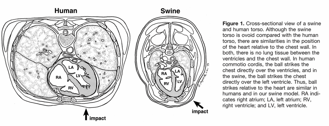
Another question is begged - I have seen pigs used for many cardiac arrest related studies, specifically for continuous compressions. Is there a specific, non-economic reason we are using pigs? Is it just that their heart, and its coverage by the lungs, are similar to that of a human?
 
I figured that it would "kick off" like an "R on T" or similar issue where the heart is electrically stimulated at a very specific part of the T wave. What was very interesting to me was the repol info you posted. That pretty much cemented in my mind how (and why) the heart ends up in VF after an event. That was pretty much one of those lightbulb moments...
 
Since you mentioned that K+ channels being blocked can prevent VF, I do have a question relating to this.
The K+ channel blocking preventing VF was in this particular instance rather than all VF.

I'm reading that pigs are similarly effected with hypo and hyperkalemia. Which makes sense, given the similar structures. Naturally, the T wave is the primary tell on a 12-lead of hyperkalemia. The fact that the impact results in R-on-T, involving the T wave, and that K+ involves the T wave can't be a coincidence, especially when blocked K+ channels reduce occurrences of VF. But this seems backwards to me. Wouldn't this indicate that K+ essentially slows down repolarization, and increases the max dispersion? Guess I need to look into the roles of each of the different channels.
I believe this was a bit less of a practical finding and more of a deduction finding. Put like so: if the K+ channels allow for VF, if we block them will VF occur. They didn't mention if it also messed up normal heart function, etc. They tried Ca2+ channel blocking (the other major channel which has an impact on electrical charge w/ mechanical stress) but that did not prevent VF. Therefore they determined it to be primarily mediated by K+. K+ blocking would certainly slow down repolarization, so I don't disagree.

So, this is purely an electrical problem, not even caused by structural abnormalities (it was an acute force that caused it). Assuming the patient is resuscitated in an effective and timely manner, is it just a "Hey I'm back" kind of resuscitation, or is there still risk for bradycardia, hypotension, or arrhythmia?
My understanding is that commotio cordis has favorable outcomes if CPR and defibrillation is initiated early.

Another question is begged - I have seen pigs used for many cardiac arrest related studies, specifically for continuous compressions. Is there a specific, non-economic reason we are using pigs? Is it just that their heart, and its coverage by the lungs, are similar to that of a human?
This is the best question so far...no idea! I've asked my partner in EMS crime Michael Herbert to answer this one. He works at a company that has a pig lab, so I reckon they'd know. I'll let you know what I find out.
 
Another question is begged - I have seen pigs used for many cardiac arrest related studies, specifically for continuous compressions. Is there a specific, non-economic reason we are using pigs? Is it just that their heart, and its coverage by the lungs, are similar to that of a human?
What I've learned is: similar respiratory rate to humans, broader chest (less likely to break ribs), less unpleasant for society than dogs.
 
What I've learned is: similar respiratory rate to humans, broader chest (less likely to break ribs), less unpleasant for society than dogs.

Thank you for your replies and the information! It is interesting that the RR is similar. Figured it was just because people don't tend to worry about pig testing.

Thanks again!
 
Another question is begged - I have seen pigs used for many cardiac arrest related studies, specifically for continuous compressions. Is there a specific, non-economic reason we are using pigs? Is it just that their heart, and its coverage by the lungs, are similar to that of a human?

(1) It's a large mammalian model that's relatively easy to get ethical permits for. Try getting approved to do the same experiments with dogs. The animal rights crowd are less likely to show up at your lab when you're working with an animal that's considered "live stock". I'm not even sure we should be allowed to do experimentation on chimpanzees and other higher primates.

(2) EP is fairly similar. Rodents are great when you want to examine ion channel functions (e.g. you can knock out pretty much whatever channel you want for ~ $100K), but hard to instrument, e.g. arterial line on a 35g mouse (can be done, but is difficult), but have heart rates of 300-600/min, a less distinct plateau phase on single-cell recording, and have a combined QRS-T complex without a distinct ST complex.

(3) Anatomic similarities.
 
Christopher said:

Brother, I've heard you explain a lot of things, and this is one of the best morsels of education I've seen yet. All that textbooking is paying off.
 
Back
Top