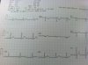
Hi! So I got an ER tech job and have been working since the start of the summer! I have some training in EKG as well and find them to be interesting. What is your opinion of this one? I am not an expert but the 12 lead appears to be fine except for aVL looks strange to me. Maybe some sort of bundle branch block but I could be totally off. Thanks!
You are using an out of date browser. It may not display this or other websites correctly.
You should upgrade or use an alternative browser.
You should upgrade or use an alternative browser.
EKG puzzler
- Thread starter jedi88
- Start date
Jambi
Forum Deputy Chief
- 1,099
- 1
- 36
Well, Lead I may have a little ST depression, but it may just be the angle, but that and aVL are contiguous. I'd like to see 5 and 6.
My first thought though are lead placement, and my second thought is it may just be an RSR presentation and not bad...but all of the conjecture is in a vacuum...
My first thought though are lead placement, and my second thought is it may just be an RSR presentation and not bad...but all of the conjecture is in a vacuum...
stemi
Forum Crew Member
- 88
- 0
- 6
It looks fine to me considering all of the other leads and axis look good. For a bundle branch block, you'd wanna focus on the precordial leads, since those will usually tell you whether you have LBBB or RBBB. As for fascicular block, axis looks good.
If anyone else has any thoughts, please chime in.
If anyone else has any thoughts, please chime in.
MSDeltaFlt
RRT/NRP
- 1,422
- 35
- 48
It's benign. What you have there is a pretty good looking 12-Lead.
sirengirl
Forum Lieutenant
- 238
- 32
- 28
It's benign. What you have there is a pretty good looking 12-Lead.
Agreed. I could see how you would think that it might be a BBB but in the absence of any other leads beig involved, and that it seems to be a normal heart rate, and the QRS doesn't appear to be widened from what I can see, I would say it's benign also.
....I should upload the picture a fellow medic sent me from the ER a few weeks back. That one was some craziness... Stand by.
sirengirl
Forum Lieutenant
- 238
- 32
- 28
Okay, so, not so much of a puzzler...
...but nonetheless impressive. Medic friend in the ER sent this to me, and then told me the patient walked in through the ER complaining of chest pain.
:huh:......noooooo, really?!


...but nonetheless impressive. Medic friend in the ER sent this to me, and then told me the patient walked in through the ER complaining of chest pain.
:huh:......noooooo, really?!
MSDeltaFlt
RRT/NRP
- 1,422
- 35
- 48
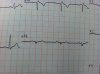
Good 12-Lead and good cath pics.. Do you know what type of AMI that ? And what artery is involved by looking at the ECG ?
sirengirl
Forum Lieutenant
- 238
- 32
- 28
Good 12-Lead and good cath pics.. Do you know what type of AMI that ? And what artery is involved by looking at the ECG ?
(you're making me think, which is unfair when I haven't been in class in 2 weeks!) That is, how you call, in das boot, inferior MI. As for the artery, I'm guessing circumflex, but I am likely wrong, my brain's been turning to moosh. Need to start studying for that state....
But yeah, he said the door-to-balloon time nearly set a record at the hospital.
MSDeltaFlt
RRT/NRP
- 1,422
- 35
- 48
Right on Inf AMI. The artery involved would be RCA. Because of the ST elevation in II, III, and aVF and also with reciprocal depression in I and aVL. ST elevation will show the general area of blockage where the reciprocal depression will show that it is on the other side making the diagnosis more accurate.
(you're making me think, which is unfair when I haven't been in class in 2 weeks!) That is, how you call, in das boot, inferior MI. As for the artery, I'm guessing circumflex, but I am likely wrong, my brain's been turning to moosh. Need to start studying for that state....
But yeah, he said the door-to-balloon time nearly set a record at the hospital.
RCA actually. Circumflex is generally is a branch of LCA, though it can originate directly from the aortic cusp.
sirengirl
Forum Lieutenant
- 238
- 32
- 28
meh, I knew I was likely wrong.
In any case, still an impressive cath picture...
In any case, still an impressive cath picture...
MSDeltaFlt
RRT/NRP
- 1,422
- 35
- 48
meh, I knew I was likely wrong.
In any case, still an impressive cath picture...
No worries. You're a student and learning. That's what this is all about.
I mentioned in another post circumflex because I recall reading that it feeds the inferior part of the left ventricle, that in a inferior myocardial infarction, there is a small chance the circumflex is the culprit. At the moment, it makes me more suspicious that it's the circumflex because the right coronary artery usually feeds the SA and AV node, no arrhythmia right now in that EKG, but of course, it also depends where's the blockage in the right coronary artery too.
In her (cath) picture though, it's clearly the right coronary artery.
In a 12-lead, are there any clues that would indicate to you that it's not the normal artery usually associated with the type of infarct?
Edit: I decided to do a quick Google. It says that if the circumflex was the culprit, usually the STE was bigger in lead II than in lead III, and vise versa if the right coronary artery was the culprit. 93% sensitivity, 100% specificity.
http://www.ncbi.nlm.nih.gov/pubmed/10624066
Just clicked the first thing. I am gonna test that right meow.
Edit: Interesting.
Last edited by a moderator: