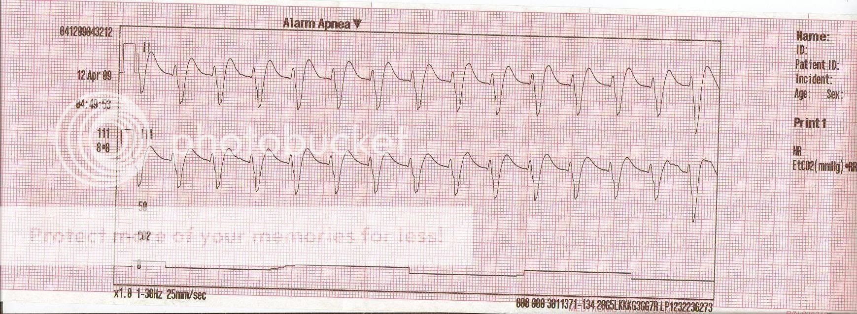
Strip please

jedirye

Forum Lieutenant
Messages
114
Reaction score
0
Points
0
strip-1.jpg


Negative on a 12-lead, as you can see other things were going down. Regardless, some sort of aberrancy?

-rye
 
Well, possibly. Is there a Lead I? With all 3 limb leads we could tell axis deviation. It is at least a wide complex tach (of unknown origin) with physiologic left axis. If Lead I is also negative, then it would be an extreme right axis making the rhythm ventricular in origin.

The axis deviation would greatly determine your treatment plan.
 
Was this a PEA (little or no CO2)?

I will go with accelerated IVR
 
I certainly agree it's wide complex. I also agree there's some flavor of axis deviation. If the patient is sick, that's one thing, but if they're chillin...I'd let axis deviation go. I may even see P waves. (Hard to tell w/o a 12 lead). Was the CO2 line hooked up? If so, then I'd say the patient had real problems.
 
Strip

Rate about 110/min. Wide. Very regular ( I mapped it out) - you'd be brave not to assume this is VT particularly in a symptomatic pt in the field. If you muddled about trying to figure it out beyond that you may a get a wake up call soon thereafter. More leads would be nice and 12lead the best of course.

It's a single focus ventricular tachycardia irrespective of aberancy or otherwise - it should be treated that way. Even an accelerated IVR should be treated likewise when the rate is over 100/min.

Sorry Delta I don't follow the "axis determining treatment option" remark.

Cheers

MM
 
Sorry Delta I don't follow the "axis determining treatment option" remark.

There are a couple of possibilities so far. VT, accelerated IVR, or even some junctional tachy rhythm with aberrant conduction. Determining the axis could put more weight on it being VT if there was ERAD (extreme right axis deviation), as it would show the mean vector of the electrical impulse originating in the ventricles in all 3 leads. This would guide the treatment of the patient both chemically and if needed electrically.

Don't even know as yet if he has a pulse yet.
 
Vt or nigh

I would make the point firstly that 2 leads is clearly a suboptimal information starting point.

However there is already a very wide QRS demonstrated so the conduction pathway is clearly non standard and the impulse vector is slowed across the ventricles. I can't see such a wide rhythm originating around the AV node to make junctional a differential interpretation.

As for treatment unless your guidelines are substantially different from mine whether arrested or with a pulse even as a PEA (perhaps especially as a PEA) the pt would get electricity +/- Ventricular anti-arrhythmic and general perfusion management.

Happy to be corrected if you think I am off the mark.

I think the general principle to consider a fast wide tachyarrhythmia as ventricular needing expeditious intervention has been borne out in practice especially with the time delays encountered in the old EMS rhythm puzzle of discerning the SVT with aberrancy from the VT as an example- this is not always possible even in the ED with 12leads. The same can apply in the field.

In one doesn't treat it as a ventricular arrhythmia then what do you treat it as? And what happens to your pt in the meantime?

I guess that depends on what you interpret this rhythm as in the first place which is of course the aim of this thread.

More information needed.

MM

PS looking at that CO2 reading (assuming it's accurate) this is more than llikely a pulseless pt.
 
Last edited by a moderator:
I can't see such a wide rhythm originating around the AV node to make junctional a differential interpretation.

Perhaps, but what if it was junctional with a BBB? Not that I think it is myself.

As for treatment unless your guidelines are substantially different from mine whether arrested or with a pulse even as a PEA (perhaps especially as a PEA) the pt would get electricity +/- Ventricular anti-arrhythmic and general perfusion management.

Interesting. Do you happen to have any links to any research that proves that shocking PEA is in of any benefit? Unless, by elecricity, you mean TCP for the brady PEA.

The way I see it, with PEA, there is usually an organized electrical pattern on the monitor, just without a palpable pulse. It could technically be any rhythm, although the common ones are slow and wide. So what is there to defibrillate? The cardiac cells can still be depolarizing in synch, just without either an associated physical contraction of the myocardium, or insufficient cardiac output to cause a viable pressure. Over here, PEA is all about H's, T's, Epi, and Atropine (if it is brady). More or less treated like asystole with some critical thinking thrown in.

I am still hesitant to call it VT, but it really is all about how the patient presented. The CO2, more than the EKG, tells me he is not well (there is a waveform and CO2 reading of 8) so I would be thinking imminent code either way.
 
Last edited by a moderator:
Interesting. Do you happen to have any links to any research that proves that shocking PEA is in of any benefit? Unless, by elecricity, you mean TCP for the brady PEA.

The way I see it, with PEA, there is usually an organized electrical pattern on the monitor, just without a palpable pulse. It could technically be any rhythm, although the common ones are slow and wide. So what is there to defibrillate? The cardiac cells can still be depolarizing in synch, just without either an associated physical contraction of the myocardium, or insufficient cardiac output to cause a viable pressure. Over here, PEA is all about H's, T's, Epi, and Atropine (if it is brady). More or less treated like asystole with some critical thinking thrown in.

Please note I said +/- electricity/antiarrhythmic and perfusion management. Our guidelines are the same by the sound of it. Bradys are treated as you describe and epi/cpr etc to start, with the option then to treat arrhythmias identified at pulse/rhthym check as per appropriate guideline. So once you've worked the perfusion angle to begin with if you have shockable rhythm then shock it - this would of course include VT with or without a pulse hence my comment.
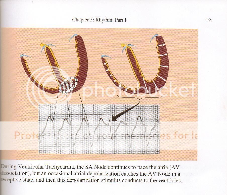
We had this very same discussion, funnily enough, in another thread just recently. It diverged into the merits or otherwise of shocking asystole (by calling it fine VF) and other topics.

I guess it depends (and it hasn't been identified by the original poster) what the clinical setting for this rhythm strip is. It may well be in the setting of an infarct, arrest - who knows. We could all end up just discussing hypotheticals - not such a bad thing and the exercise of examining the rhythm is always worthwhile don't you think?

MM

PS We also have an accelerated IVR guideline with a <60/min, 60-100/min and > 100/min rate approach- the greater than 100/min is to treat an AIVR as VT - so the pt would get shocked (if arrested or with extreme poor perfusion)
 
Last edited by a moderator:
Let's not make a mountain out of a molehill. This is why we have more leads (even on our most basic monitors we have three leads, not two...). Assumping the capnography tracing is accurate, you'd have to assume the pt is in a pulseless VT and proceed with the ACLS assault:deadhorse:. If the patient had a pulse, I'd lean away from VT, frankly. I still haven't ruled out the presence of well-behaved P waves (which would make this a ST with some kind of BBB )...and we'd look silly trying to convert that. Need. More. Data.
 
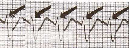
At the risk of some sort of copyright infringement (all credit to Dr. Dubin), but I came across some information while reading...

book.jpg


StripwithArrows.jpg


smallversionstrip.jpg
 
At the risk of some sort of copyright infringement (all credit to Dr. Dubin), but I came across some information while reading...
book.jpg


StripwithArrows.jpg


smallversionstrip.jpg

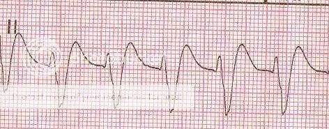
Thanks for the comparison ECG - it really throws a spanner into the works or does it?

I don't believe this rhythm shows any characteristics of a bundle branch block pattern such as an rSR profile as seen in RBB. (Of course we still don't have the right leads to make a definitive judgement on that).

Also note in the comparison of the strips that the whilst looking similar they are in fact different. Half way down the S wave of the original strip there is a notched change in the slope perhaps identifying a p wave - hence this may be considered AV dissociation if it is, a characteristic necessary to lean towards VT.

In the end the dilemma remains, if you saw this and the pt was crook what would you do? I for one would still have to lean towards treatment of VT as I find it unlikely a pt would be symptomatic with a Sinus tach with BBB (or pulseless and arrested as is likely here given that tantalising CO2 reading).

Whilst we can't make a definitive interpretation one way or the other based on the level of info I find the exercise of pulling the rhythm to bits to see what makes it tick very helpful. Tneds to weed out my old bad habits of putting things in baskets.

Certainly interested in other opinions of course.

MM
 
Last edited by a moderator:
Back
Top