EKG question

squirrel15

Forum Captain
299
144
43
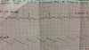
I'm an EMT-B that just started working on CCT unit, and want to learn how to read EKG's so I have a better understanding what is actually happening when I'm in the back of the rig. We mostly use 3 leads and occasionally 12 leads. Unfortunately the nurse I'm with isn't much of the teaching type. She gave me an old strip and told me to figure out what's wrong with it over the weekend. Only info I have is it was on a three lead.

At first look I was thinking afib, but considering afib is an irregular heartbeat, could this just be artifact? Because to me, it seems like the actual heartbeat is regular, since the qrs waves are spaced apart quite evenly, there is just no p or t wave.

Thanks for the help!
 

Attachments

  • 2015-02-22 22.14.30.jpg
    2015-02-22 22.14.30.jpg
    1.9 MB · Views: 232

STXmedic

Forum Burnout
Premium Member
5,018
1,356
113
You're correct in thinking it's just artifact. There are visible P waves, and the QRSs are evenly spaced. Sinus Tach is the rhythm you're looking at.

As to you learning, I'll make he same suggestion I make whenever this question comes up (which is damn near once a week). If you want to teach yourself ECG interpretation, read:

Walraven's Basic Arrhythmias
then
Dubin's Rapid Interpretation of ECGs
then (if you still want more)
Garcia's 12 Lead Art of Interpretation

After you get the depth you want, read 12 leads. Daily. The more you read, the better you'll get. If you google WaveMaven, they have a huge library of 12 leads to peruse through.

However, at the EMT level, it's all just nice to know. Other than making appropriate destination choices, it doesn't really change how you function.
 
OP
OP
S

squirrel15

Forum Captain
299
144
43
Ok thanks! Yeah, I know it doesn't change how I function, but sometimes its nice to know what you're looking at and not always needing something explained to you. I'll look in to getting those books though, and I'm hoping to get partnered with a more energetic nurse in the near future too that is more willing to answer dumb questions :)
 

Sunburn

Forum Crew Member
30
7
8
That BPM is around 100 so its borderline tach. What you got there is ST depression, which is indicative of myocardium in trouble.
 

Aprz

The New Beach Medic
3,031
664
113
That BPM is around 100 so its borderline tach. What you got there is ST depression, which is indicative of myocardium in trouble.
In monitor mode, tachycardia, and in a single lead only, I wouldn't worry too much about ST depression.

ST Changes in Monitor Mode

I don't completely understand filtering, but what I do know is that monitor mode can: create ST elevation that isn't there, ST depression that isn't there, or mask ST changes that are occurring. On the Philips HeartStart MRx, which is the same monitor the OP is using, you have to do a 12-lead to view the leads in diagnostic mode.

You can read more about filtering at ems12lead.com.

In my limited experience, it's not very common for the 3-lead mode ST change to be different from what I'd see on a 12-lead, but it does happen every once in awhile. If I see ST changes in 3-lead mode or if the patient has a complaint that warrants a 12-lead even though I don't see ST elevation in monitor mode, I am gonna do a 12-lead.

ST Depression in Tachycardia

You probably know about the QRS complex representing the ventricles depolarizating and the T-wave represents the ventricles repolarizing. The p-wave presents the atria depolarizing, right? What about the atria depolarizing? At a normal heart rate, the atria repolarize about the same time as the ventricles depolarizing and the ventricles are way bigger than the atria so it is hidden within the QRS complex. In tachycardia though, you can sometimes see slight ST depression because the atria are repolarizing.

Using a Single Lead

There are many things that cause ST changes. On emsbasics.com, Brandon Oto shared some stats about ST elevation and chest pain. In patients with chest pain AND ST changes, only 15% of them were a STEMI. Other causes for the ST changes were left ventricular hypertrophy (#1 cause, more than MI), left bundle branch block (equal to number of MI patients), benign early repolarization, ventricular paced rhythm, ventricular aneurysm, and pericarditis. By looking at a single lead alone, how can you attribute the ST changes to ischemia?
 

Sunburn

Forum Crew Member
30
7
8
Yeah, I'd very much like to see 12 lead. Sure you would not dg nSTEMI from a single II lead, but that is the only single problem with that ECG tracing. His BP is a tad high too which is consistent with partial blockage and/or myocardial distress. I'd like to see the patient and 12-lead for sure. With what we have it's just a guess.
 

STXmedic

Forum Burnout
Premium Member
5,018
1,356
113
Yeah, I'd very much like to see 12 lead. Sure you would not dg nSTEMI from a single II lead, but that is the only single problem with that ECG tracing. His BP is a tad high too which is consistent with partial blockage and/or myocardial distress. I'd like to see the patient and 12-lead for sure. With what we have it's just a guess.
It's also consistent with high blood pressure.... Probably much more so than an MI...

3 lead strip and single lead, the only thing "wrong" is tachy. There's nothing else legitimate (that's not a stretch and tremendous "well maybe what if") you can pull from that monitor-view strip.
 

Sunburn

Forum Crew Member
30
7
8
Exactly. Diagnosing anything from a single lead is pointless. It's a blind men describing an elephant problem.
But the question was not to find diagnosis, it was to find what is not normal with that strip, and the answer to that is solved :p
 

chaz90

Community Leader
Community Leader
2,735
1,272
113
Since it's a single lead, I would guess the nurse's point in asking him to find out what was "wrong" in this strip was just to illustrate simple sinus tach. Perhaps she's trying the whole "keep looking for zebras that aren't there" approach as she shows him what is, overall, a fairly normal and common EKG.
 
OP
OP
S

squirrel15

Forum Captain
299
144
43
So I found out what the problem was, and it does not show in this picture sorry! But it wasn't a long enough strip and didn't have an spo2 reading on it. That was all.
 

Aprz

The New Beach Medic
3,031
664
113
So I found out what the problem was, and it does not show in this picture sorry! But it wasn't a long enough strip and didn't have an spo2 reading on it. That was all.
Wow, that is ultra lame. To be honest, that nurse kinda sounds like a snob now. I avoid working with people like that at all cost because they are usually "the problem". I would not consider the lack of the SpO2 being printed on the strip a problem at all. People usually write down what they see while they are looking at the monitor at that moment or send over the data to their ePCRs if possible. If you somehow missed the SpO2, you can print out trends if the monitor hasn't been turned off or print out the trends via data management if you have turned it off.

I used to work CCT a lot and nurses like that made me sick of CCT. I felt like a lot of them wanted to work with me because I was pretty experienced (for CCT IFT as an EMT) and knowledgeable about this kinda stuff, but I didn't want to work with them because of things like that. Ridiculous. I almost went back to working on BLS, lol, but then I got hired on with ALS 911.
 
Last edited:

DieselBolus

Forum Crew Member
80
33
18
My guess is she wasnt looking for an interp, she probably wanted you to say that there was too much road noise and deviation in the iso line. Lead placement could probably fix that. Nothing better than when someone places leads ON a skinny pts clavicles.
 
Top