EKG Interp/scenario take 2

Really?

Perhaps I'm misunderstanding what you've written -- but it sounds like you're saying all of your patients with post-arrest VT have had coronary occlusions on angiography? Is this published anywhere? How many patients is 100%?

no what I am saying is that if the initial rhythm is VF/VT and we get pulese back they go to the cath lab (by protocol). it is a small couldn't so there aren't any published studys, yet. we had 25 saves of 78 arrests with initial rhythm VF/VT, all 25 had stints placed in the cath lab.
 
Those are clearly PVC's. I wouldn't even waste my time trying to vagal them down with a HR of 120 (not that you would even want to), which I'm assuming the PVC's are perfusing ones. I'd immediately get an IV and hang a bag of 150 mg of Amiodarone over 10 min and get them going to a hospital.
 
no what I am saying is that if the initial rhythm is VF/VT and we get pulese back they go to the cath lab (by protocol). it is a small couldn't so there aren't any published studys, yet. we had 25 saves of 78 arrests with initial rhythm VF/VT, all 25 had stints placed in the cath lab.

AHA Policy Statement: Regional Systems of Care for Out-of-Hospital Cardiac Arrest

"Up to 71% of patients with cardiac arrest have coronary artery disease, and nearly half have an acute coronary occlusion.48–50 There is a high incidence (97%) of coronary artery disease in patients resuscitated from OOHCA who undergo immediate angiography and a 50% incidence of acute coronary occlusion.48 However, the absence of ST elevation on a surface 12-lead electrocardiogram after resuscitation of circulation from cardiac arrest is not strongly predictive of the absence of coronary occlusion on acute angiography.48 A case series of patients with unsuccessful field resuscitation suggested that in such patients, VF is more likely to be due to coronary disease than is asystole or pulseless electric activity.51 [...] these studies suggest that patients who are resuscitated from out-of-hospital VF have a high likelihood of having an acute coronary occlusion."
 
thanks tom
 
time for a new one

83 yr old male AMS

5.jpg
 
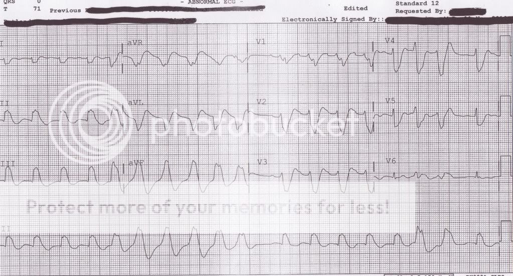
here we go 72 yr old female c/c CP ems is bls. 12 lead is done transmitted shows ST elevation II,III, avf with reciprocal changes laterally, 5 min from hospital pt has arrest cpr and defib x1 this is the 12 lead as shes being rolled in the ED

31.jpg
First EKG V-tach with ST elevations. Solution ACLS
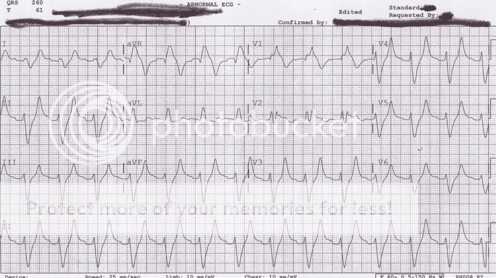
Second EKG V-fib. Solution CPR, call coroner.
 
First ECG looks like some sort of wicked bundle branch block or hyperkalemia

Second is either diagnostic mode VF or it's VT

If its VT Brown would cardiovert first and ask questions or hang amiodarone later
LOL
Brown why can't I get your stains out of my boxer.
lol all in good fun.
 
There's something you shouldn't see - a 12 lead of VF.
 
maby the 12 lead was already on when he went into v-tach. i would not have taken the time to do more than the std 4 lead in a symptomatic VT. he would have been riding the lightning, edison medicine, aka synchronized cardioversion. then prophylactic lidocaine or amio.
 
45.

It is most likely supraventricular rhytm + LBBBB. Maybe hyperkalemia, maybe MI.

If he has chest pain/stenocardia, you should treat him like MI.
Of sublinqual Nitro doesnt stop pain:
-Nitroglycerin IV
-Morphine
-Ac.sal. acid
-Metoprolol or Amiodarone
 
Last edited by a moderator:
Back
Top