ECG Cold Read Challenge

Christopher

Forum Deputy Chief
1,344
74
48
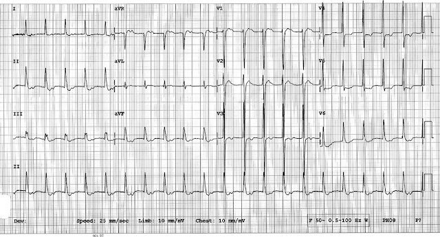
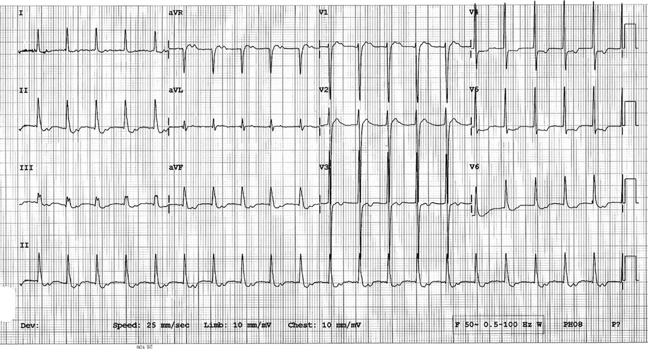
(These ECG's are from a paper I was reading and represent a good interpretation and diagnosis to have locked away in your memory)

66 year old female, ECG #1 from the outlying hospital. ECG #2 is from after transfer at a larger academic hospital.

No history, no nothing, because everybody loves a cold read!


Summit

Critical Crazy
2,693
1,314
113
I'll try... my 12 lead skills are weak and I'm working on them.

JT or regular afib rvr
increasing inferior and lateral ST depression, possible septal wall MI but more likely subendocardial injury
incomplete developing block intrafasicular block

In for some better interps.
 

tchristifulli

Forum Crew Member
82
0
0
Sinus tach with a 1st degree av block. Axis is normal. Some sort of IVCD but not a BBB yet. Left ventricular hypertrophy. Anterior and septal depression. Is she on Digoxin?
 

Wes

Forum Lieutenant
193
0
16
Seems too regular for A-Fib. I'm seeing some patterns that look like possible digoxin toxicity as well. What about WPW?
 
OP
OP
Christopher

Christopher

Forum Deputy Chief
1,344
74
48
At least I can't see P wave. Is it not a sign of A-fib?

It is best to be specific when talking about the presence or absence of P-waves.

What you really want to ask: is there discernible atrial activity? P-waves? F-waves? f-waves? (F-: flutter, f-: fib)

This is where a close examination of the TP interval and QT interval are most important. The TP interval is typically where you see evidence of atrial activity, like P-waves or F-/f-waves.

You also can see atrial activity in the QT interval depending on the conditions, because the atria marches to its own beat (in most cases).

So before giving up hope of P-waves (or F-/f-waves) check out the QT interval too!
 
OP
OP
Christopher

Christopher

Forum Deputy Chief
1,344
74
48
Seems too regular for A-Fib. I'm seeing some patterns that look like possible digoxin toxicity as well.

Regularized atrial fibrillation is sometimes seen in digoxin toxicity. Although, a close inspection of the QT/TP intervals shows no evidence of f-waves, so I would agree that regularized afib is not likely.

What about WPW?

No evidence of preexcitation present and given the rate it seems unlikely to be orthodromic AVRT. Provided this lady was on some healthy doses of meds which slowed a bypass tract...that could be the case.

I think given any supraventricular tachycardia with secondary ST/T-wave changes it is wise to consider the possibility of an accessory pathway, just at the patient's age and the tachycardia's rate I wouldn't put it high on my list.
 

Medic496

Show strength when weak, and humility when strong
59
4
8
Cold Read

My best guess is what is to ask what the blood pressure and pulse ox is. If stable, transport to someone smarter than me.
 

Aprz

The New Beach Medic
3,031
664
113
My best guess is what is to ask what the blood pressure and pulse ox is. If stable, transport to someone smarter than me.
It's a cold read so you don't get any of that information. At minimum, try breaking it down. Maybe that'll help you figure it out. :)

What's the rate?

What's the rhythm?
-- Is it regular?
-- Are there p-waves present?
-- Are the p-waves associated with the complexes?
-- What's the PR interval?
-- Are the complexes wide?

What's the QTc? (To calculate QTc, it's QT/((60/HR)^(1/2)).)

What's the mean QRS axis?

What about the axis of the precordial lead? (ie is there normal R-wave progression?)

Is there any hypertrophy?

Any Q-waves?

Any retrograde T-waves?

Any ST depression or elevation? If so, what are they shaped?

^^This is usually how I break down my 12-lead.

Even if you don't get the answer right, at least you've broken down the ECG, and you'll recognize characteristics of the problem next time when you see it because you've broken it down into little bits of detail.
 
Last edited by a moderator:

Summit

Critical Crazy
2,693
1,314
113
0.30 buried in T

I see what you are looking at, but that would make for an inverted p wave (negative deflections in II and V leads, positive deflection in AVR). That doesn't happen except in junctional rhythms (and then you don't have a long pri) or weird ectopic atrial rhythms.
 

tchristifulli

Forum Crew Member
82
0
0
I see what you are looking at, but that would make for an inverted p wave (negative deflections in II and V leads, positive deflection in AVR). That doesn't happen except in junctional rhythms (and then you don't have a long pri) or weird ectopic atrial rhythms.

ImageUploadedByTapatalk1375547191.968549.jpg
It's not inverted
 
OP
OP
Christopher

Christopher

Forum Deputy Chief
1,344
74
48
And the answer is?

The first ECG represents a junctional tachycardia with retrograde capture of the atria. Another possibility to consider is regularized atrial fibrillation, however, there is no evidence of any f-waves. Flat/Scooped ST-segment depression is present.

The second ECG represents either a slower junctional tachycardia or a sinus tachycardia with a quite prolonged PRi. A cleaner strip would help. Scooped ST-segment depression is clearly present.

In either case, this ECG is pathognomonic for Digitalis Toxicity.

In this case the patient had normal dig levels and normal K+ levels, however, they had critically low magnesium levels which caused an acquired digitalis toxicity. Treatment of the patient's mag levels solved their arrhythmia.

I only bring this up as it is worth keeping in your head the common causes for each type of arrhythmia. We usually stress interpretation, but less so Dr. Garcia's "Consider the Company it Keeps".

If you see Junction Tachycardia or Regularized Atrial Fibrillation your first thought should be Digitalis.

(once you announce this somebody will inevitably chime in that you can't give Calcium...you can promptly ignore them)
 
Top